Focus on Electrophysiology | His Bundle Pacing: A More Physiologic Alternative For Pacing
Believe it or not, we're still debating the optimal site for pacing the ventricle. Despite our success in treating bradyarrhythmias, we're continually looking for better ways to treat our patients, improve outcomes and decrease long-term complications.
Electrical and mechanical dyssynchrony secondary to chronic right ventricular (RV) pacing can result in long-term deleterious effects. Two trials (DAVID and MOST) demonstrated an increased risk of hospitalization for heart failure (HF) in patients having a high burden of RV pacing and consequently an increased risk of arrhythmias.1,2
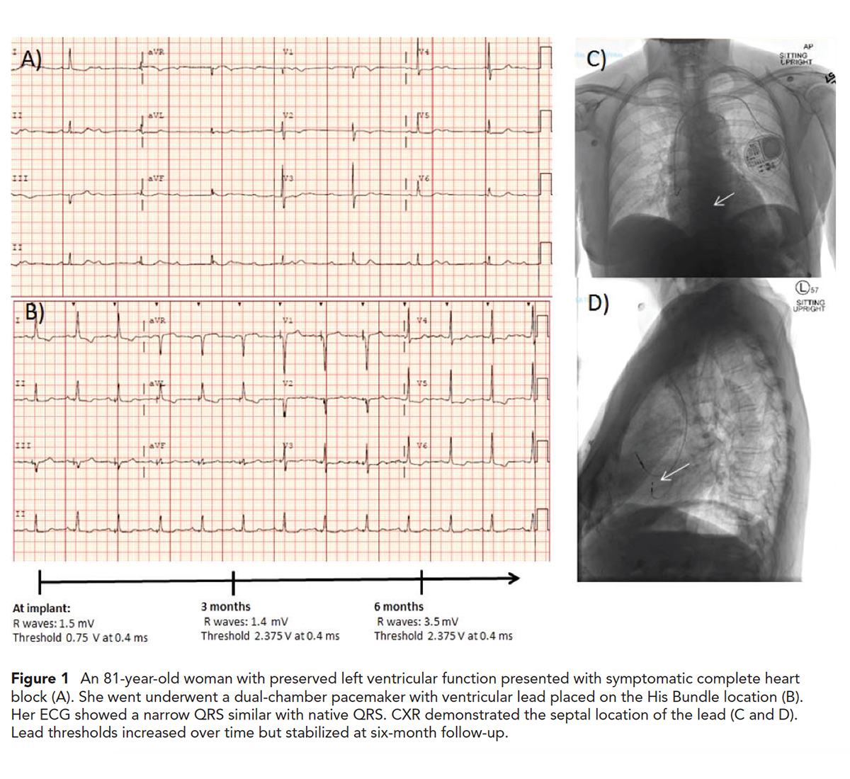
 Click the image above for a larger view.
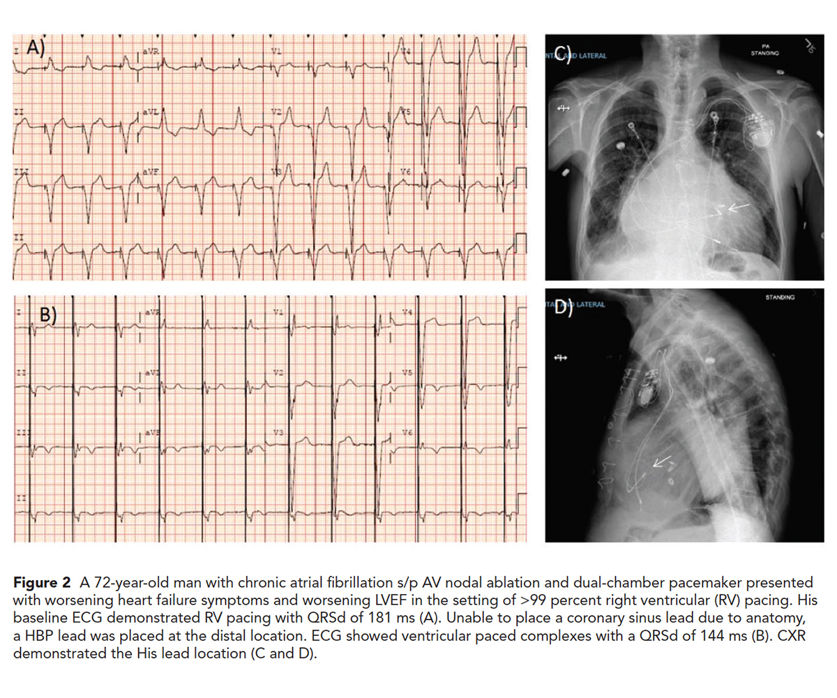
 Click the image above for a larger view.
 Click the image above for a larger view.
The potentially harmful effects of long-term RV pacing may occur in patients with either preserved and reduced left ventricular (LV) systolic function. However, patients with a reduced left ventricular ejection fraction (LVEF) at baseline suffer more consequences.
Multiple strategies have been explored to combat this problem and minimize the RV pacing burden: new pacing algorithms, alternative RV pacing sites as well as cardiac resynchronization therapy (CRT).
Prior studies have explored other alternatives to traditional apical RV pacing, such as RV septum and RV outflow tract. However, there's no clear evidence of superiority of these sites compared with traditional RV apical pacing. In addition, data suggest that worsening of LVEF occurs with any RV pacing site.3
Another strategy to minimize RV pacing is using algorithms to limit the amount of RV pacing when possible. These algorithms use long atrioventricular (AV) delays, which may impair AV synchrony and can potentially lead to an increased risk of AV block at higher atrial rates and predispose to mitral regurgitation in certain patients.4 Therefore, patient selection is important.
On the other hand, the benefits of CRT over RV pacing are well established in patients with LV dysfunction, left bundle branch block (LBBB) with long QRS duration and high-degree AV block. However, we still have a group of selected patients with preserved LV function for whom the indication of CRT remains controversial.
His bundle pacing (HBP) offers a more physiologic alternative mode of pacing the ventricle using the native His-Purkinje system. While a physiologically and logically appealing alternative, the practical approach can be challenging.
Placement of the lead and achieving stability can be technically difficult. It is important to emphasize that this idea is not new. It was first described almost two decades ago by Deshmukh and colleagues.5 In the last decade numerous case series have been published demonstrating the feasibility and utility of HBP in clinical settings.6
There are numerous challenges to this technique. Having a solid understanding of the anatomy is key. The His bundle is an isolated structure, a continuation of the AV node that provides a connection for the electrical impulse to travel from the AV node to the right and left ventricle via the right and left bundles, respectively. There are multiple variations in anatomy of the bundles, which add more complexity to the procedure and its success. Initially HBP was performed using regular pacing leads, which were not designed with the unique anatomic characteristics of the His bundle taken into account. This made the procedure challenging and time consuming.
The development of new tools such as the SelectSecure 3830 (Medtronic, Minneapolis, MN) and sheath (C315His, C304 SelecSite) have made these procedures more widely used. However, the tools are still limited, especially for patients with an enlarged right atrium and/or displaced tricuspid annulus.
Further, there is concern about maintaining chronic pacing therapy due to the risk of lead dislodgement, exit block and concern about progressive electrical block distal to the HBP lead. However, more recently, with increased procedural experience, the feasibility of permanent HBP, including patients with infranodal AV block, has been demonstrated to be >90 percent. In addition, recent studies suggest similar fluoroscopy and procedural times compared with RV pacing.7 Nevertheless, while there have been many published case studies and single-center reports of HBP, there have been no large randomized clinical trials.
A new meta-analysis of 26 papers of permanent HBP showed an overall average implant success rate of 84.8 percent. From baseline to last follow-up, there was an average 5.9 percent increase in LVEF with statistical significance (p=0.001), with a greater increase among patients with a history of HF and correspondingly lower baseline LVEF.7
Terminology is also important. What does HBP means? Recently, a multicenter HBP Collaborative Working Group proposed a refined set of criteria to define HBP in patients with normal His-Purkinje conduction and in those with His-Purkinje conduction disease.8 The authors defined two forms of His bundle capture: selective capture, in which the His bundle is the only tissue captured by the pacing stimulus; and nonselective capture, in which there is fusion capture of the His bundle and adjacent ventricular tissues.
Early reports of HBP are very promising. However, many questions still remain, especially in patients with severe distal conduction system disease. Some authors have reported good results using a distal location in the His-bundle axis. This may allow for locating the pacing site beyond the site of conduction block in patients with intra-Hisian AV block or bundle branch block. This distal location often produces a nonselective His-bundle capture with better pacing thresholds and ventricular sensing parameters, thus it may eliminate the need for backup ventricular pacing lead.9
HBP has grown in popularity over the last few years. The recent guideline on bradycardia and conduction delay from the ACC/American Heart Association gave a Class IIa indication for patients with AV block who have an indication for permanent pacing with an LVEF between 36-50 percent and who are expected to require ventricular pacing more than 40 percent of the time.10 A Class IIb indication was given for patients with AV block at the level of the AV node who have an indication for permanent pacing.
However, at this stage there isn't sufficient evidence to perform HBP instead of CRT in patients with depressed LVEF and LBBB. A coronary sinus lead continues to be the preferred treatment for these patients until more data become available. HBP should be used as a bail-out when a LV lead placement is not possible.
Two large randomized clinical trials are underway to helps us clarify the role of HBP in pacing for patients with LBBB, RBBB, prolonged PR intervals, or an expected high degree of ventricular pacing in the setting of underlying HF. One is called the His-SYNC, comparing HBP and coronary sinus pacing for CRT. The other is called the HOPE-HF trial and is evaluating the optimal pacing strategy in HF.
There is no doubt that HBP is a safe and feasible approach to pacing with physiologic advantages. However, further research and innovation from industry is necessary before HBP can become more widely used as the pacing mode of choice. The excellent implant success rate and lead parameters reported by experienced operators need to be replicated globally. Large randomized clinical studies with long-term data are needed for this technology to move forward.

This article was authored by Eliany Mejía-López, MD, a fellow in training in the Cardiac Electrophysiology program at the University of Virginia Medical Center, Charlottesville, VA. She thanks Rohit Malhotra, MD, FACC, who provided insights and expertise that greatly assisted the work and cases reviewed here.
References
- Wilkoff BL, Cook JR, Epstein AE, et al. D JAMA 2002;288:3115-23.
- Lamas GA, Lee KL, Sweeney MO, et al. N Engl J Med 2002;346:1854-62.
- Beck H, Curtis AB. Curr Heart Fail Rep 2016;13:230-6.
- Lewis AJM, Foley P, Whinnett Z, et al. J Am Heart Assoc 2019;8:e010972.
- Deshmukh P, Casavant DA, Romanyshyn M, Anderson K. Circulation 2000;101:869-77.
- Sharma PS, Dandamudi G, Naperkowski, A et al. Heart Rhythm 2015;12:305-12.
- Zanon F, Ellenbogen KA, Dandamudi G, et al. Europace 2018;20:1819-26.
- Vijayaraman P, Dandamudi G, Zanon F, et al. Heart Rhythm 2018;15:460-8.
- Vijayaraman P, Chung MK, Dandamudi G, et al. J Am Coll Cardiol 2018;72:927-47.
- Kusumoto FM, Schoenfeld MH, Barrett C, et al. J Am Coll Cardiol 2018;Oct 31:[Epub ahead of print].
Keywords: ACC Publications, Cardiology Magazine, Electrophysiology, Bundle-Branch Block, Cardiac Resynchronization Therapy, Bundle of His, Atrioventricular Block, Atrioventricular Node, Bradycardia, Mitral Valve Insufficiency, Heart Ventricles, Coronary Sinus, American Heart Association, Patient Selection, Stroke Volume, Atrial Fibrillation, Follow-Up Studies, Ventricular Function, Left, Heart Failure, Heart Atria, Hospitalization, Fluoroscopy, Algorithms
< Back to Listings