An 80-year-old Caucasian man with a history of hypertension presented with syncope. An electrocardiograph (ECG) demonstrated third-degree atrioventricular (AV) block and a non-contrast transthoracic echocardiogram showed a left ventricular (LV) ejection fraction of 35% with severe hypokinesis of the LV apex. A dual-chamber pacemaker was implanted. For further evaluation of his decreased ejection fraction, a nuclear perfusion study was performed that showed a small fixed perfusion defect in the inferior wall, although a diaphragmatic artifact could not be ruled out. Cardiac catheterization was subsequently performed to evaluate for ischemic heart disease. There was no evidence of obstructive coronary disease, and left ventriculography was performed as shown (Figure 1, Video 1).
Figure 1
Video 1
Which of the following statements explains these findings?
Show Answer
The correct answer is: D. Apical variant hypertrophic cardiomyopathy.
Apical variant hypertrophic cardiomyopathy (HCM), also known as Yamaguchi Syndrome, is seen in only 1 to 2% of HCM cases in the non-Japanese population. Characteristically, there is a narrow LV cavity at the apex with preserved dimensions at the base, resembling a "spade" shape (Figure 2). The pigtail catheter in this image is seen in the mid LV cavity and contrast material appears dark and completely fills the LV cavity, except in the apical section, due to cavitary obliteration by the thickened myocardium. Note the hyperdynamic LV function. Occasionally, non-contrast enhanced echocardiographic images may be misinterpreted as having a low LV function due to poor visualization of the endocardial borders at the apex. Apical aneurysm of the LV is seen as ballooning and outpouching of the LV wall contour. LV apical thrombus is visualized as a localized intracavitary radiolucency with a "moth-eaten" appearance, often seen in the presence of low ejection fraction.
Figure 2
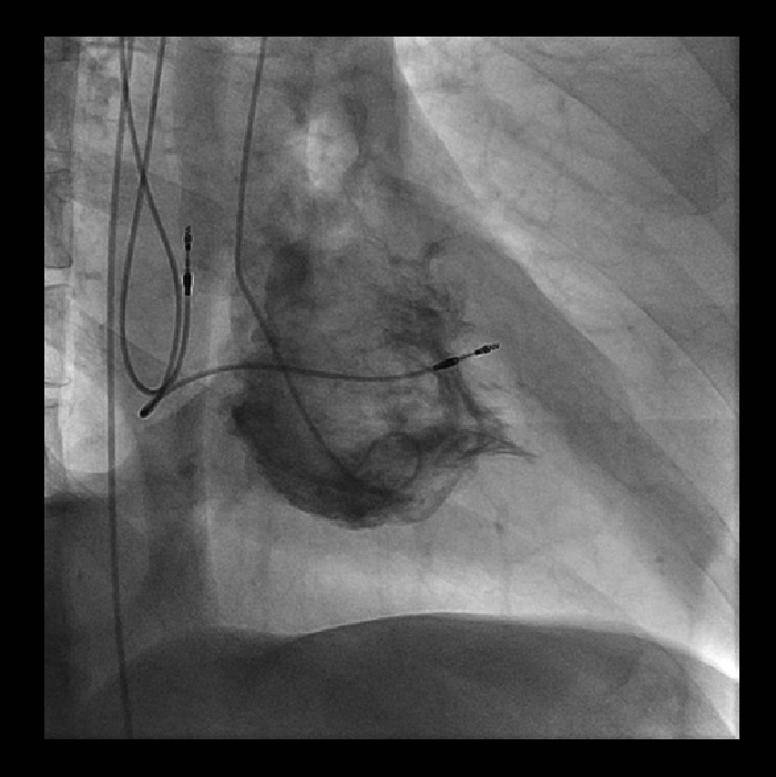
Left ventriculography in the right anterior oblique (RAO) projection demonstrating a "spade"-shaped left ventricular (LV) cavity, suggestive of apical variant hypertrophic cardiomyopathy (HCM). Contrast agent appears dark and defines the narrow channel-like apical cavity (white arrow). At the apex, there is increased myocardial thickness (black arrows). Also seen are the dual-chamber pacemaker leads (asterisks).
References
Zoffoli G, Mangino D, Venturini A, et al. Diagnosing left ventricular aneurysm from pseudo-aneurysm: a case report and a review in literature. J Cardiothorac Surg 2009;4:11.
Kimura K, Tanabe-Hayashi Y, Noma S, Fukuda K. Images in cardiovascular medicine. Rapid formation of left ventricular giant thrombus with Takotsubo cardiomyopathy. Circulation 2007;115:e620-1.
Otieno H, Vivas Y, Traub D, Raman A, Polam C. Images in cardiovascular medicine: contrast echocardiography in apical hypertrophic cardiomyopathy. Circulation 2006;114:e33-4.