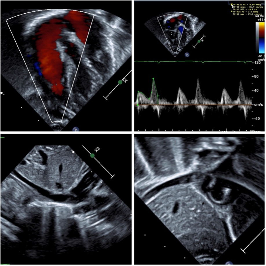
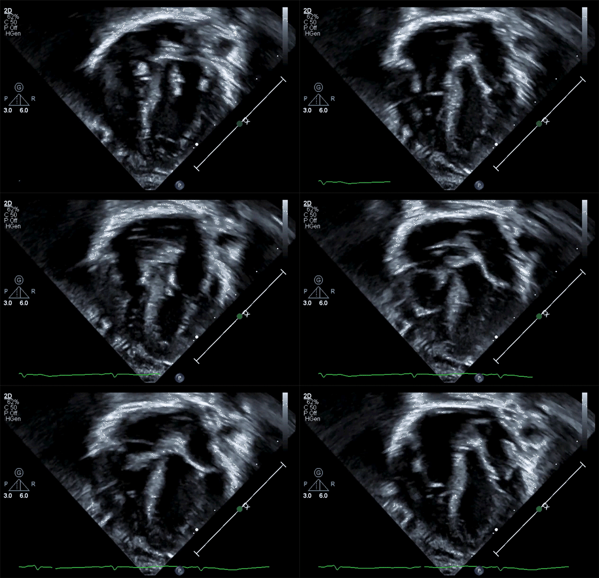
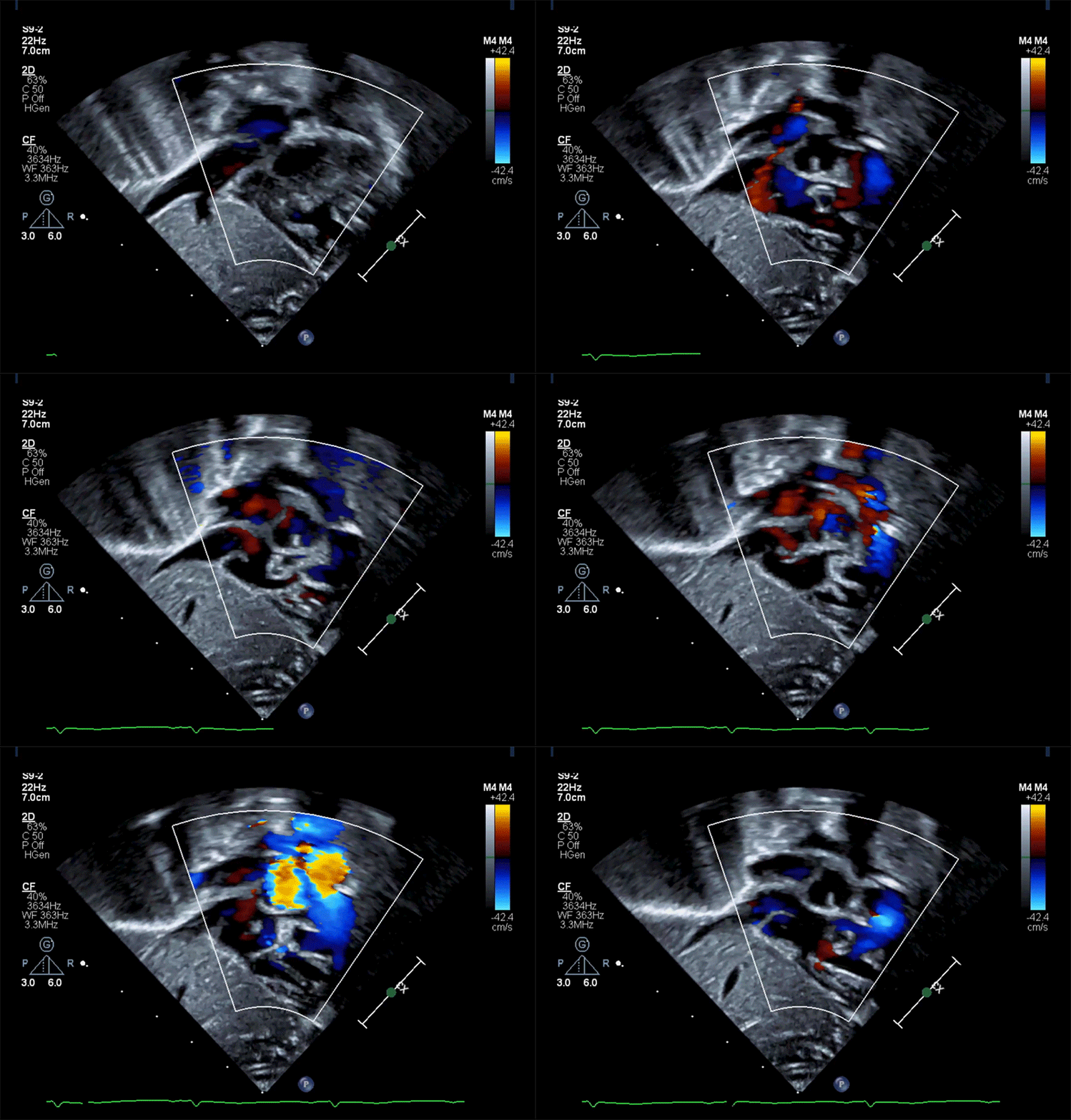
This patient's echocardiogram demonstrated normal tricuspid valve hinge points, normal flow through the tricuspid valve, and normal-sized intracardiac structures and chambers. Panel A in Figure 2 shows unobstructed tricuspid valve flow (white arrow). Panel B shows a normal tricuspid valve Doppler pattern and velocity. Panel C shows a subcostal view of the inferior vena cava draining into the right atrium. The eustachian valve is visualized (white arrow), which is separate from the mobile membranous structure in the right atrium seen in Videos 1 and 2. Panel D shows a bicaval view with a mobile echogenic structure in the right atrium (white arrow), separate from the eustachian valve (asterisk). The appearance of this mobile structure is consistent with a prominent Chiari network.
Figure 2: TTE at Presentation
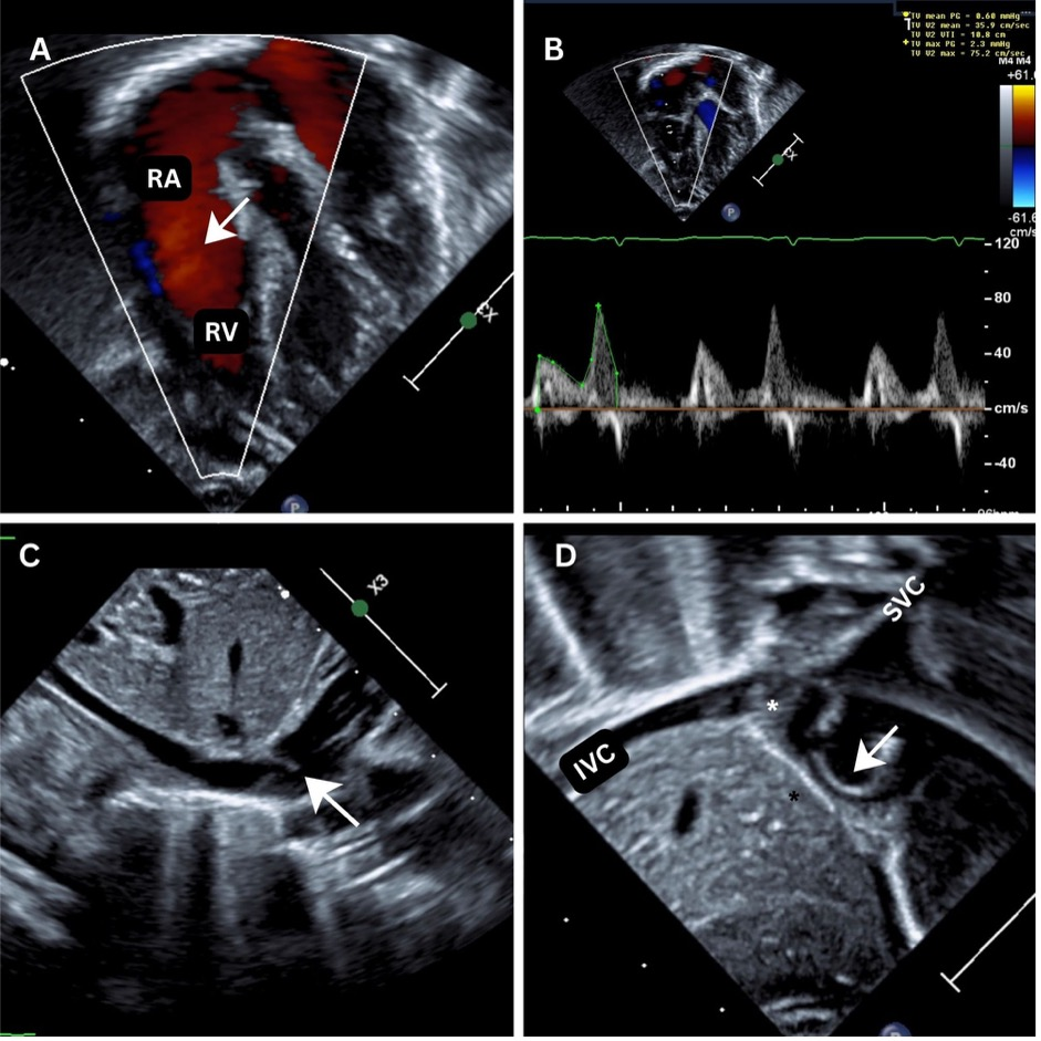
IVC = inferior vena cava; RA = right atrium; RV = right ventricle; SVC = superior vena cava; TTE = transthoracic echocardiogram.
A Chiari network, present in approximately 2% of the population, is a reticulated network of fibers in the right atrium resulting from the incomplete involution of the right valve of the sinus venosus and can be discovered as an incidental finding on diagnostic imaging studies.1 It is a thin, highly mobile curvilinear structure that moves with blood flow within the right atrium.2 Other embryologic remnants such as a prominent eustachian valve (valve of the inferior vena cava) and crista terminalis can also appear as echogenic structures in the right atrium and mimic right atrial masses.3 On echocardiography, the crista terminalis appears as a fixed, well-defined fibromuscular ridge with echogenicity similar to the adjacent myocardium, located along the posterolateral right atrial wall near the superior vena cava. The eustachian valve is a thin, leaf-like linear structure arising from the inferior vena cava–right atrial junction.
Echocardiography can also help differentiate these benign embryologic remnants from pathological findings such as tumors, vegetations, and thrombi. A thrombus usually appears as an echodense mass with margins distinct from the endocardium. In this patient, the echocardiographic characteristics were unlike a right atrial thrombus. The clinical history and echocardiographic findings were not consistent with a vegetation, which can appear as oscillating or filamentous masses attached to intracardiac catheters, valve structures, or endocardial surfaces. Therefore, an evaluation or management for endocarditis with blood cultures and antibiotics would not be warranted.
Although there are rare cases of transient neonatal cyanosis associated with a Chiari network partially obstructing the tricuspid valve,4 her echocardiographic findings showed normal tricuspid valve flow and a left-to-right atrial-level shunt. Thus, the Chiari network was considered an incidental finding. In this patient, the intermittent desaturations were thought to reflect persistent fetal circulation (i.e., intermittent right-to-left atrial-level shunting due to transient elevation in pulmonary vascular resistance). Her cyanotic episodes resolved after 2 days, supporting this diagnosis.
In summary, in a stable patient with self-resolving intermittent desaturations and an incidental finding of a Chiari network causing no clinical symptoms or tricuspid valve obstruction, no intervention or follow-up is needed. The intermittent desaturations may be related to persistent fetal circulation and should be managed conservatively.
References
- Joyce JJ, Joyce TR, Ferns SJ. Prominent prolapsing Chiari network: presentation and prognosis in paediatric patients. Cardiol Young. 2022;32(7):1071-1076. doi:10.1017/S1047951121003668
- Kim MJ, Jung HO. Anatomic variants mimicking pathology on echocardiography: differential diagnosis. J Cardiovasc Ultrasound. 2013;21(3):103-112. doi:10.4250/jcu.2013.21.3.103
- Trento A, Zuberbuhler JR, Anderson RH, Park SC, Siewers RD. Divided right atrium (prominence of the eustachian and thebesian valves). J Thorac Cardiovasc Surg. 1988;96(3):457-463.
- Aljemmali S, Bokowski J, Morales R, Abdulla RI. Chiari network associated with hypoxemia in a neonate: case report and review of the literature. Pediatr Cardiol. 2020;41(7):1529-1531. doi:10.1007/s00246-020-02432-w