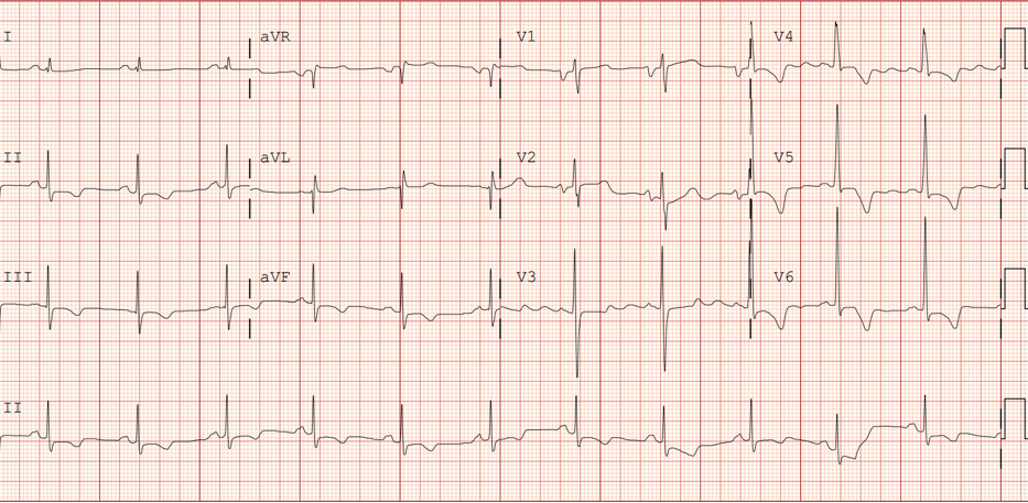
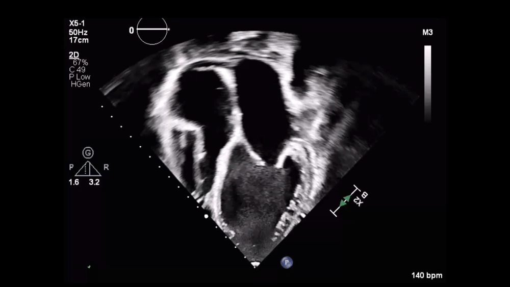
This patient has symptomatic heart failure (New York Heart Association class II and Ross class II symptoms) with reduced EF (HFrEF) due to dilated cardiomyopathy. She is currently treated with a diuretic and a beta-blocker but previously discontinued an angiotensin-converting enzyme (ACE) inhibitor due to cough.
Optimization of renin-angiotensin-aldosterone system (RAAS) inhibition remains a core component of GDMT in pediatric HFrEF. The 2025 International Society for Heart and Lung Transplantation (ISHLT) Guidelines for the Management of Pediatric Heart Failure recommend angiotensin receptor-neprilysin inhibitor (ARNI) therapy as a reasonable alternative to single agent ACE inhibitor or angiotensin-receptor blocker (ARB) in children >1 year of age with symptomatic systemic LV systolic dysfunction (Class 2a, Level of Evidence B).1
This recommendation is based on substantial evidence of efficacy in adults with HFrEF and the PANORAMA-HF (Prospective Trial to Assess the Angiotensin Receptor Blocker Neprilysin Inhibitor LCZ696 Versus Angiotensin-Converting Enzyme Inhibitor for the Medical Treatment of Pediatric HF) trial, which compared sacubitril/valsartan with enalapril in 375 children with systemic LV systolic dysfunction. Although sacubitril/valsartan did not demonstrate superiority over enalapril in the primary global rank endpoint at 52 weeks, it showed comparable clinical improvement and an acceptable safety profile, leading to approval by the United States Food and Drug Administration for use in children >1 year of age with symptomatic LV systolic dysfunction.1,2 In this patient who did not tolerate an ACE inhibitor, transitioning to an ARNI/ARB combination is the most appropriate next step in GDMT (answer choice B).
This patient developed a cough following initiation of an ACE inhibitor. ACE inhibitor-induced cough is a well-described side effect of ACE inhibitors and is mediated by the accumulation of bradykinin. Angioedema is a rarer but more significant side effect associated with ACE inhibitors and is also associated with bradykinin accumulation. ARBs have a much lower risk of cough because this class of medications does not inhibit bradykinin degradation.3,4 In this patient, switching to an ARNI combination medication such as sacubitril/valsartan is a reasonable, evidence-based alternative that is preferable to restarting enalapril at a lower dose (answer choice A).
Ivabradine (answer choice C) is reasonable adjunctive therapy for pediatric patients with chronic systolic HF. In children with dilated cardiomyopathy, elevated heart rate has been associated with increased risk of death and cardiac transplantation.5 According to the 2025 ISHLT Guidelines for the Management of Pediatric Heart Failure, ivabradine is a reasonable agent to use in children with stable HF when heart rate reduction is desirable (Class 2a, Level of Evidence B).1 This patient's heart rate is 65 bpm on carvedilol therapy, which is low-normal for her age; thus, ivabradine is not the best next step in medical therapy for this patient.
Vericiguat (answer choice D) is a soluble guanylate cyclase stimulator that promotes vasodilation, improves myocardial function, and reduces adverse cardiac remodeling. In adults with HFrEF, vericiguat has been studied as an adjunctive therapy in patients receiving GDMT. The VICTOR (Vericiguat Global Study in Participants With Chronic Heart Failure) trial, a large, randomized, placebo-controlled study of >6,000 adults with HFrEF, showed a significantly lower incidence of cardiovascular death among patients receiving vericiguat over a median follow-up of 18.5 months.6 A phase 2/3 clinical trial is currently underway to evaluate the safety and efficacy of vericiguat in pediatric patients with HF. Vericiguat may be a reasonable addition to GDMT once all other medications have been optimized.1 However, this is not the most appropriate next step in therapy for this patient at this time because GDMT has not yet been fully implemented.
In summary, RAAS inhibition remains a crucial component of GDMT in pediatric HFrEF. According to the new ISHLT Guidelines for the Management of Pediatric Heart Failure published in 2025, ARNI therapy is a reasonable alternative to an ACE inhibitor or ARB in children >1 year of age with symptomatic systemic LV systolic dysfunction. The PANORAMA-HF trial demonstrated safety and comparable efficacy of ARNI to enalapril in pediatric patients. GDMT optimization should precede consideration of adjunctive agents.
References
- Irving C, Azeka E, Adorisio R, et al. The International Society for Heart and Lung Transplantation guidelines for the management of pediatric heart failure (update from 2014). J Heart Lung Transplant. 2025;44(10):e21-e71. doi:10.1016/j.healun.2025.06.003
- Shaddy R, Burch M, Kantor PF, et al. Sacubitril/valsartan in pediatric heart failure (PANORAMA-HF): a randomized, multicenter, double-blind trial. Circulation. 2024;150(22):1756-1766. doi:10.1161/CIRCULATIONAHA.123.066605
- Borghi C, Cicero AF, Agnoletti D, Fiorini G. Pathophysiology of cough with angiotensin-converting enzyme inhibitors: how to explain within-class differences?. Eur J Intern Med. 2023;110:10-15. doi:10.1016/j.ejim.2023.01.005
- Lochbaum R, Hoffmann TK, Greve J, Hahn J. Concomitant medication in patients with bradykinin-mediated angioedema – there's more than ACE inhibitors. J Dtsch Dermatol Ges. 2023;21(11):1283-1289. doi:10.1111/ddg.15154
- Bonnet D, Berger F, Jokinen E, Kantor PF, Daubeney PEF. Ivabradine in children with dilated cardiomyopathy and symptomatic chronic heart failure. J Am Coll Cardiol. 2017;70(10):1262-1272. doi:10.1016/j.jacc.2017.07.725
- Butler J, McMullan CJ, Anstrom KJ, et al. Vericiguat in patients with chronic heart failure and reduced ejection fraction (VICTOR): a double-blind, placebo-controlled, randomised, phase 3 trial. Lancet. 2025;406(10510):1341-1350. doi:10.1016/S0140-6736(25)01665-4