Exercise-Induced VT in a Former Cross-Country Runner
A 25-year-old male runner with no significant medical history presents for evaluation of exertional palpitations and presyncope, with frequent ventricular ectopy noted on electrocardiogram (ECG) (Image 1). He was a former competitive track and cross-country runner through high school and college, training at high intensity with reported times as fast as 4:30 min/mile. He reports a 6-month history of progressive exertional palpitations, lightheadedness, and dyspnea. His symptoms culminated during a 5K race while recovering from a viral upper respiratory illness lasting 3-4 days, when he developed abrupt palpitations, diaphoresis, severe dyspnea, and near-syncope. Similar episodes subsequently recurred with exertion. He denies stimulant or performance-enhancing drug use. His family history is notable for sudden death with exertion in a maternal great-uncle.
Image 1
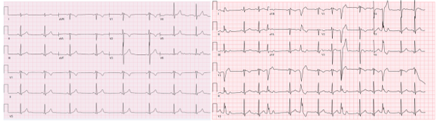
ECG from the patient's initial presentation with findings notable for baseline NSR, frequent monomorphic ventricular ectopy, and an rSr′ pattern in lead V1 with fractionation suggesting late activation.
ECG = electrocardiogram; NSR = normal sinus rhythm.
A transthoracic echocardiogram reveals normal left ventricular (LV) size and systolic function (ejection fraction 56%), no LV hypertrophy (maximal wall thickness 1 cm), and borderline abnormal global longitudinal strain (-15.9%). The right ventricle (RV) appears qualitatively enlarged, with right atrial size at the upper limit of normal. No significant valvular abnormalities are present.
A 48-hour Holter monitor reveals a predominant rhythm of sinus bradycardia (mean 53 bpm) with frequent ventricular ectopy: multifocal isolated premature ventricular complexes (PVCs; 2.2% burden), couplets, triplets, runs of nonsustained ventricular tachycardia (VT), and episodes of accelerated idioventricular rhythm (Image 2).
Image 2
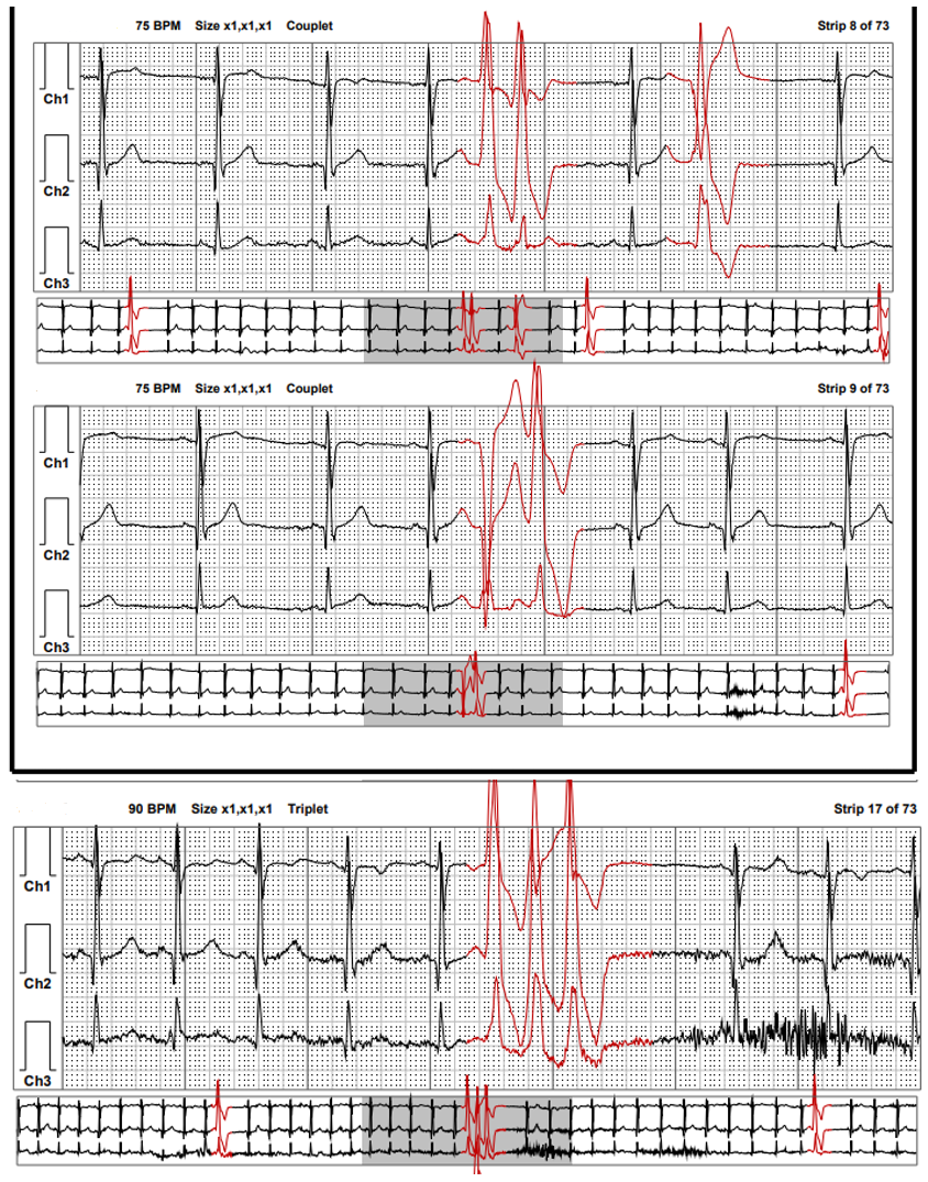
Representative 48-hour ambulatory ECG monitoring tracings demonstrating frequent ventricular ectopy.
ECG = electrocardiogram.
Given concern for frequent multifocal PVCs and exertional symptoms, he undergoes a treadmill ECG stress test. Early during exercise, he has incessant runs of repetitive pleiomorphic VT (Image 3).
Image 3
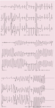
ECG tracing from the patient's stress test with findings notable for the development of PMVT.
ECG = electrocardiogram; PMVT = polymorphic ventricular tachycardia.
Despite intravenous diltiazem, esmolol, and amiodarone, he continues to experience recurrent VT. Because he remains hemodynamically stable throughout and the episodes briefly self-terminate, electrical cardioversion is not required. Owing to the persistent arrhythmia burden, he ultimately undergoes endotracheal intubation with fentanyl and propofol, resulting in suppression of VT and maintenance of sinus rhythm. He is subsequently admitted to the cardiac intensive care unit. Findings on initial laboratory evaluation, including electrolyte, kidney and thyroid function, and cardiac biomarker values, is unrevealing.
A cardiac MRI demonstrates normal LV size, function, wall motion, and wall thickness; normal indexed RV volumes with low-normal to mildly reduced systolic function (RV ejection fraction 41%); and no evidence of RV trabeculations, focal aneurysms, or dyskinesia (Videos 4a, b, c). There is no late gadolinium enhancement or evidence of fatty infiltration. A PET-CT demonstrates no myocardial uptake suggestive of active inflammation. A coronary CT angiogram shows normal coronary artery origins without coronary artery disease.
Video 4a
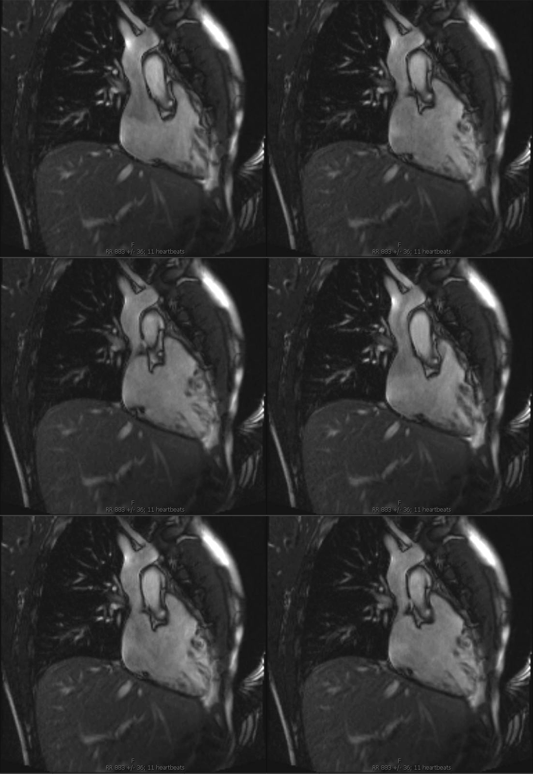
Cardiac MRI of the RV inflow tract demonstrating normal function and no evidence of increased RV trabeculations, focal aneurysms, or dyskinesia.
RV = right ventricular.
Video 4b
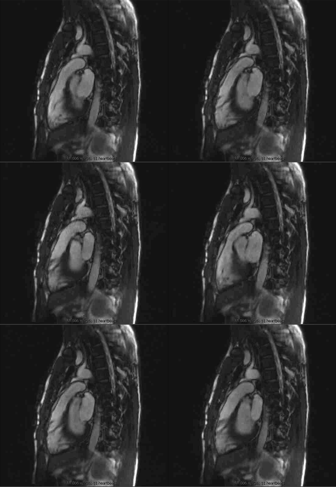
Cardiac MRI of the RV outflow tract demonstrating normal function and no evidence of increased RV trabeculations, focal aneurysms, or dyskinesia.
RV = right ventricular.
Video 4c
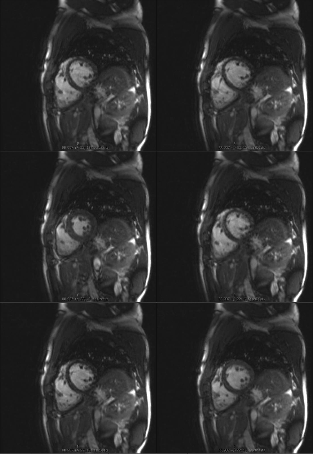
Cardiac MRI SAX view of the RV demonstrating normal function and no evidence of increased RV trabeculations, focal aneurysms, or dyskinesia.
RV = right ventricular; SAX = short-axis.
Which one of the following is the best next step in his management?
Show Answer


