Clinical Practice Algorithm For the Follow-up of Unrepaired and Repaired Secundum Atrial Septal Defects
Quick Takes
- Small secundum atrial septal defects (ASDs) do not require frequent surveillance and many close spontaneously in early childhood.
- Moderate and large secundum ASDs should be closed when they cause right ventricular volume overload, generally during the preschool years to prevent long-term complications.
- After surgical or transcatheter ASD closure, long-term follow-up can be spaced out to 2-5 years if there are no significant residual lesions or clinical concerns.
Background
There is lack of evidence-based guidelines to help cardiologists determine the frequency of follow-up and testing in children with congenital heart disease (CHD). Thus, there remains a wide variation in clinical practice even for the management of common types of CHD.1 The CHD Clinical Practice Algorithms are designed to provide clinicians with a decision-support tool for management of CHDs, both before and after catheterization- or operation-based interventions. This algorithm is the first of a series of clinical practice algorithms that are focused on isolated secundum atrial septal defects (ASDs).
Goals and Details of the Algorithm
The goal of the ASD clinical practice algorithm is to provide cardiologists and other health care providers with a decision-support tool for clinical evaluation and follow-up testing of secundum ASDs (Figures 1a, b, c). This guidance is not meant to replace health care providers' clinical judgment but rather to improve the value of care by reducing practice variation and optimizing resource utilization.
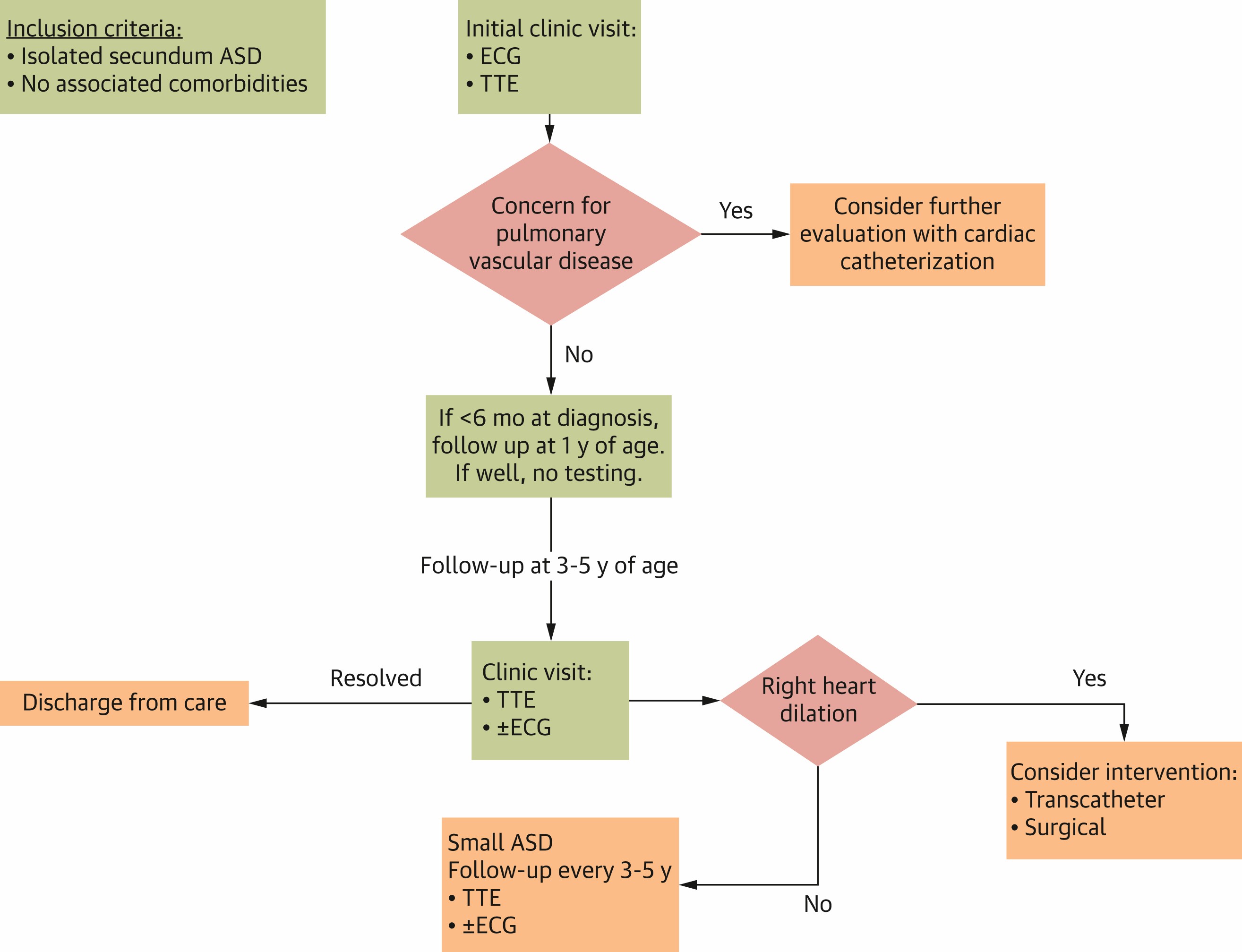
Figure 1a: Outpatient Management of Isolated Secundum ASD in Patients <5 Years of Age
Reprinted with permission from Writing Committee, Sachdeva R, Parthiban A, et al. Outpatient management of isolated left-to-right shunt lesions in pediatric patients: 2026 ACC concise clinical guidance: a report of the American College of Cardiology Solution Set Oversight Committee. J Am Coll Cardiol. Published online March 26, 2026. doi:10.1016/j.jacc.2025.11.020
ASD = atrial septal defect; ECG = electrocardiogram; TTE = transthoracic echocardiography.
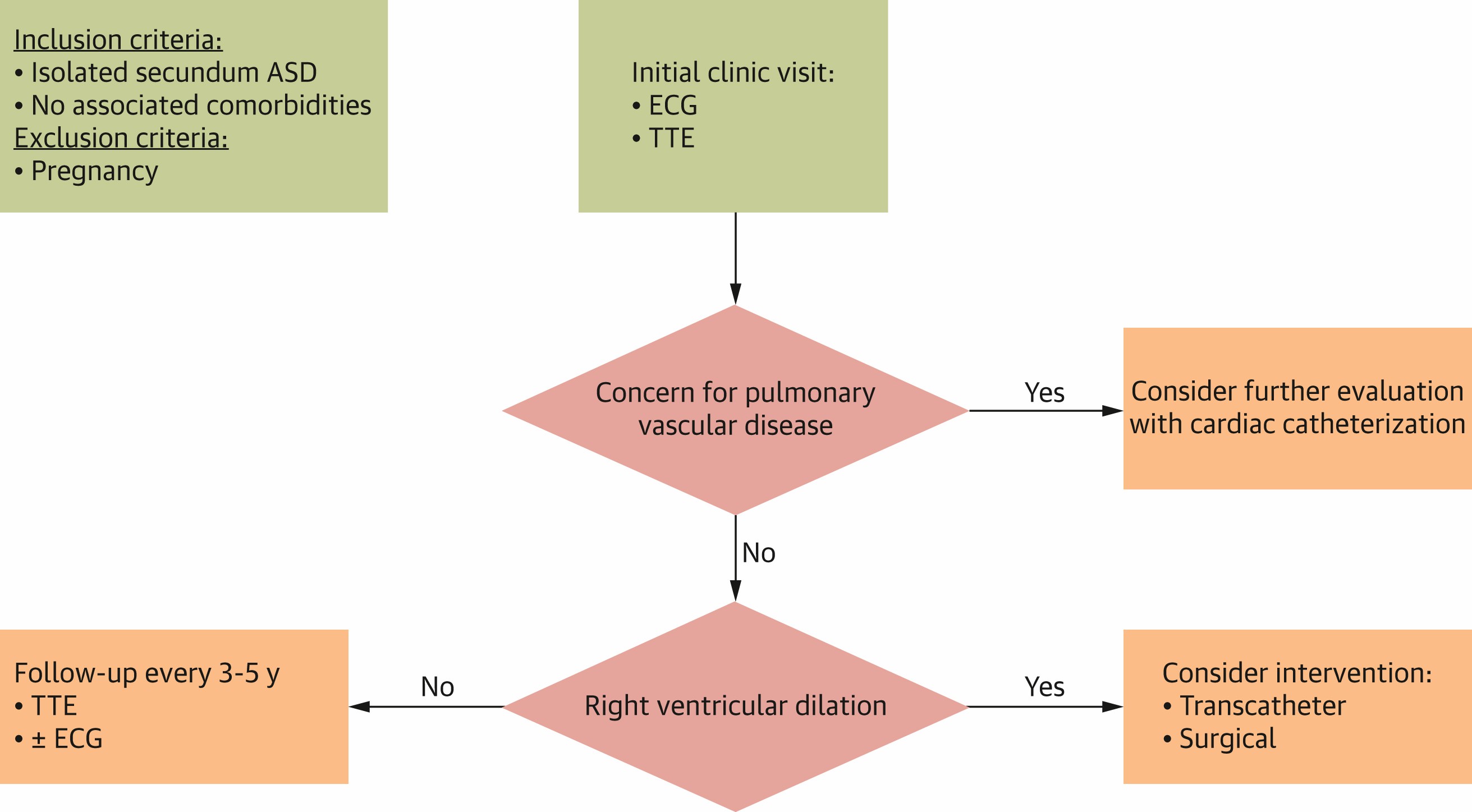
Figure 1b: Outpatient Management of Isolated Secundum ASD in Patients 5-18 Years of Age
Reprinted with permission from Writing Committee, Sachdeva R, Parthiban A, et al. Outpatient management of isolated left-to-right shunt lesions in pediatric patients: 2026 ACC concise clinical guidance: a report of the American College of Cardiology Solution Set Oversight Committee. J Am Coll Cardiol. Published online March 26, 2026. doi:10.1016/j.jacc.2025.11.020
ASD = atrial septal defect; ECG = electrocardiogram; TTE = transthoracic echocardiography.
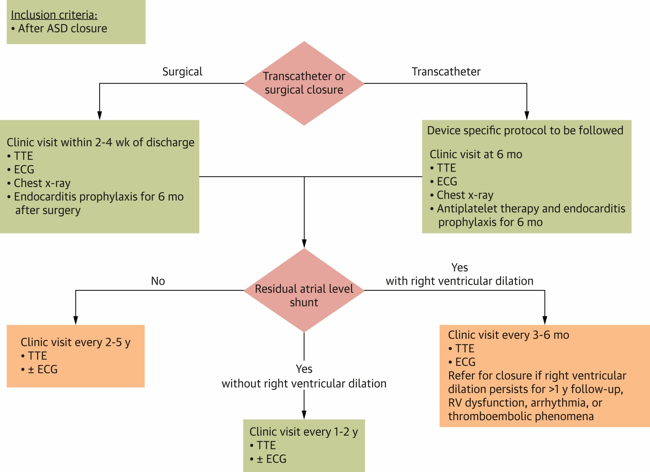
Figure 1c: Outpatient Management of Secundum ASD Post Intervention
Reprinted with permission from Writing Committee, Sachdeva R, Parthiban A, et al. Outpatient management of isolated left-to-right shunt lesions in pediatric patients: 2026 ACC concise clinical guidance: a report of the American College of Cardiology Solution Set Oversight Committee. J Am Coll Cardiol. Published online March 26, 2026. doi:10.1016/j.jacc.2025.11.020
ASD = atrial septal defect; ECG = electrocardiogram; RV = right ventricle; TTE = transthoracic echocardiography.
ASDs are one of the most common forms of CHD, with secundum ASDs being the most common type, accounting for 75% of ASDs. This algorithm excludes other types of ASDs (sinus venosus, primum, and coronary sinus) given their association with more complex anatomy. Patent foramen ovale is a remnant of fetal circulation that may be present in as many as 25% of healthy adults, is typically considered benign in childhood, and is not addressed in the algorithm. Patients with significant noncardiac comorbidities including prematurity and genetic syndromes are also excluded. Despite how frequently they are encountered, there is significant variation in the management of secundum ASDs, largely because of a lack of evidence-based guidelines.2
The algorithm provides the outpatient management of secundum ASDs before intervention by age (<5 years, ≥5-18 years) and after intervention. The management of secundum ASDs is largely based on size of the defect and its hemodynamic consequences. Small secundum ASDs are not hemodynamically significant, have a high rate of spontaneous closure when diagnosed in infancy, and can be followed every 3-5 years to reassess for right ventricular (RV) dilation by transthoracic echocardiogram.3,4 Patients and/or their families should be counseled on the risk of thromboembolic events and increased risk of decompression sickness with deep sea diving, which is low but real.5 Moderate or large secundum ASDs cause right heart dilation, and it is generally accepted that they should be closed during preschool age (~3-5 years) to prevent late complications.6 As infants and young children with even large ASDs are typically free of symptoms, annual testing with electrocardiograms and echocardiograms is not needed and could be performed prior to intervention at 3-5 years of age.
Indications for ASD closure include a hemodynamically significant shunt with Qp:Qs (ratio of pulmonary blood flow to system blood flow) >1.5:1 and/or RV dilation. For secundum ASDs that meet indications for intervention, it can be performed surgically or via transcatheter device closure. Anatomical features of the ASD, including the size of the defect and associated rims, will determine whether catheter-based intervention can be performed. The less-invasive option of transcatheter device closure is generally preferred when the anatomy is favorable, and shared decision-making with the patient and/or their family should be pursued.7
After intervention, there is still a need for periodic long-term surveillance, although it can be spaced out to every 2-5 years depending on any clinical concerns. Late complications are rare but exist, including atrial arrhythmias, pulmonary vascular disease, and persistent RV dilation/dysfunction. Bacterial endocarditis is rare after both surgical and device closure, but antibiotic prophylaxis is recommended for the first 6 months until endothelization of the device/patch material has occurred.8
(For management of unrepaired ASDs in patients >18 years of age, refer to the 2025 ACC/AHA/Multisociety Guideline for the Management of Adults With CHD.9)
Methods: Algorithm Development
The algorithm was created by the Quality Working Group of the American College of Cardiology (ACC) Adult Congenital and Pediatric Cardiology (ACPC) member section. This is a multidisciplinary group of pediatric and adult congenital cardiologists from diverse training and practice backgrounds. The algorithm is evidence based, with lesion-specific references and existing guidelines used when applicable.2-8,10 A quality-driven approach was used to translate the current evidence into best practice recommendations. Rigorous quality-improvement methodology was used, including appropriate prework to identify desired outcomes, process measures, potential unintended consequences, and required stakeholders.11,12 The algorithm was subsequently peer-reviewed for consensus.
Future Directions
The ASD clinical practice algorithm can be integrated by clinicians as a decision-support tool in their practices. Furthermore, individual centers can use them for quality-improvement initiatives. The widespread use and implementation of the ASD algorithm could improve resource utilization and standardize the care of patients with ASDs.
References
- Hokanson JS, Ring K, Zhang X. A survey of pediatric cardiologists regarding non-emergent echocardiographic findings in asymptomatic newborns. Pediatr Cardiol. 2022;43(4):837-843. doi:10.1007/s00246-021-02795-8
- Ziebell DS, Ghaleb S, Anderson J, Statile CJ. Resource utilisation in paediatric patients with secundum atrial septal defects. Cardiol Young. 2020;30(3):383-387. doi:10.1017/S104795112000013X
- Radzik D, Davignon A, van Doesburg N, Fournier A, Marchand T, Ducharme G. Predictive factors for spontaneous closure of atrial septal defects diagnosed in the first 3 months of life. J Am Coll Cardiol. 1993;22(3):851-853. doi:10.1016/0735-1097(93)90202-c
- Sachdeva R, Valente AM, Armstrong AK, et al. ACC/AHA/ASE/HRS/ISACHD/SCAI/SCCT/SCMR/SOPE 2020 appropriate use criteria for multimodality imaging during the follow-up care of patients with congenital heart disease: a report of the American College of Cardiology Solution Set Oversight Committee and Appropriate Use Criteria Task Force, American Heart Association, American Society of Echocardiography, Heart Rhythm Society, International Society for Adult Congenital Heart Disease, Society for Cardiovascular Angiography and Interventions, Society of Cardiovascular Computed Tomography, Society for Cardiovascular Magnetic Resonance, and Society of Pediatric Echocardiography. J Am Coll Cardiol. 2020;75(6):657-703. doi:10.1016/j.jacc.2019.10.002
- Meissner I, Whisnant JP, Khandheria BK, et al. Prevalence of potential risk factors for stroke assessed by transesophageal echocardiography and carotid ultrasonography: the SPARC study. Stroke Prevention: Assessment of Risk in a Community. Mayo Clin Proc. 1999;74(9):862-869. doi:10.4065/74.9.862
- Campbell M. Natural history of atrial septal defect. Br Heart J. 1970;32(6):820-826. doi:10.1136/hrt.32.6.820
- Jalal Z, Hascoët S, Gronier C, et al. Long-term outcomes after percutaneous closure of ostium secundum atrial septal defect in the young: a nationwide cohort study. JACC Cardiovasc Interv. 2018;11(8):795-804. doi:10.1016/j.jcin.2018.01.262
- Wilson W, Taubert KA, Gewitz M, et al. Prevention of infective endocarditis: guidelines from the American Heart Association: a guideline from the American Heart Association Rheumatic Fever, Endocarditis, and Kawasaki Disease Committee, Council on Cardiovascular Disease in the Young, and the Council on Clinical Cardiology, Council on Cardiovascular Surgery and Anesthesia, and the Quality of Care and Outcomes Research Interdisciplinary Working Group. Circulation. 2007;116(15):1736-1754. doi:10.1161/CIRCULATIONAHA.106.183095
- Gurvitz M, Krieger EV, Fuller S, et al. 2025 ACC/AHA/HRS/ISACHD/SCAI guideline for the management of adults with congenital heart disease: a report of the American College of Cardiology/American Heart Association Joint Committee on Clinical Practice Guidelines. J Am Coll Cardiol. 2026;87(7):822-976. doi:10.1016/j.jacc.2025.09.006
- Writing Committee, Sachdeva R, Parthiban A, et al. Outpatient management of isolated left-to-right shunt lesions in pediatric patients: 2026 ACC concise clinical guidance: a report of the American College of Cardiology Solution Set Oversight Committee. J Am Coll Cardiol. Published online March 26, 2026. doi:10.1016/j.jacc.2025.11.020
- Anderson J, Verghese G, Lihn S, et al. Quality Improvement in Congenital Heart Disease. In: Wernovsky G, Anderson RH, Kumar K, Mussatto KA, Redington AN, Tweddell JS, Tretter JT, eds. Anderson's Pediatric Cardiology. 4th ed. Elsevier; 2020.
- Friedman KG, Rathod RH, Farias M, et al. Resource utilization after introduction of a standardized clinical assessment and management plan. Congenit Heart Dis. 2010;5(4):374-381. doi:10.1111/j.1747-0803.2010.00434.x
Clinical Topics: Cardiac Surgery, Congenital Heart Disease and Pediatric Cardiology, Valvular Heart Disease, Aortic Surgery, Cardiac Surgery and CHD and Pediatrics, Cardiac Surgery and VHD, Congenital Heart Disease, CHD and Pediatrics and Quality Improvement
Keywords: Aortic Coarctation, Ductus Arteriosus, Patent, Foramen Ovale, Patent, Process Assessment, Health Care, Quality Improvement, Standard of Care, Heart Defects, Congenital, Heart Septal Defects, Ventricular, Pulmonary Valve Stenosis, Algorithms, Evidence-Based Practice, Patient Care Team, Diagnostic Techniques and Procedures, Catheterization, Certification
< Back to Listings