
AI-Enabled CCTA Reveals Premature Plaque Burden in South Asians
Quick Takes
- Among South Asian adults, 63% of study patients had coronary plaque detected by artificial Intelligence-enabled quantitative coronary plaque analysis, with nearly one-half of those aged 40-54 years exhibiting some form of plaque.
- Noncalcified plaque was the dominant subtype across all age groups, making up approximately 86% of the total plaque composition.
- Men had a significantly higher prevalence of any plaque compared with women. However, women saw a sharp catch-up increase in plaque volume after age 65 years.
Commentary based on Satish P, Vajramani A, Gupta S, et al. Age and sex specific coronary plaque characteristics among South Asians: Insights from the DILWALE CTA study. Am J Prev Cardiol. 2026;29:101567. Published 2026 Mar 18. doi:10.1016/j.ajpc.2026.1
Introduction
South Asians (SA) in the United States carry a higher burden of atherosclerotic cardiovascular disease (ASCVD) than other ethnic groups, and standard risk prediction tools often fail to capture this elevated risk.2 SA ethnicity is recognized as a risk-enhancing factor in the 2026 ACC/AHA/Multisociety Guideline on the Management of Dyslipidemia. When cardiovascular risk remains uncertain, biomarkers (lipoprotein[a] and high-sensitivity C-reactive protein) and coronary artery calcium (CAC) testing can be used to further stratify risk.2,3
An important limitation of CAC testing is the potential to underestimate overall plaque burden because it does not detect noncalcified plaque (NCP). As a result, the 2026 dyslipidemia guideline suggests that selective use of coronary computed tomography angiography (CCTA) may be considered among individuals who may have a higher burden of NCP. In the DILWALE CTA study, Satish et al. confront the gap in risk assessment among SA by using artificial intelligence-enabled quantitative coronary plaque analysis (AI-QCPA) to characterize age- and sex-specific plaque burden among SA adults living in the United States.1
Summary of the Study
The DILWALE CTA study is an electronic health record-based retrospective registry of adult SA patients within the Baylor Scott and White Health system. A total of 341 patients (median age 54 years, 31% women) were included. A high prevalence of traditional risk factors was found overall: hyperlipidemia (82%), hypertension (60%), and diabetes mellitus (32%); 63% were on aspirin therapy and 57% on statin therapy.
Overall, 63% of this SA cohort had detectable coronary plaque by AI-QCPA. As expected, younger individuals showed a greater NCP predominance, whereas calcified plaque volumes increased with age. Older age and male sex were the strongest independent predictors of both the presence of any plaque and low-attenuation plaque (LAP), a component of the noncalcified group that may represent a necrotic core that is more prone to rupture. Key findings of the study are shown in Figure 1.
Figure 1: Calcified and Noncalcified Median Plaque Volumes (in mm3) by Age Group and Sex
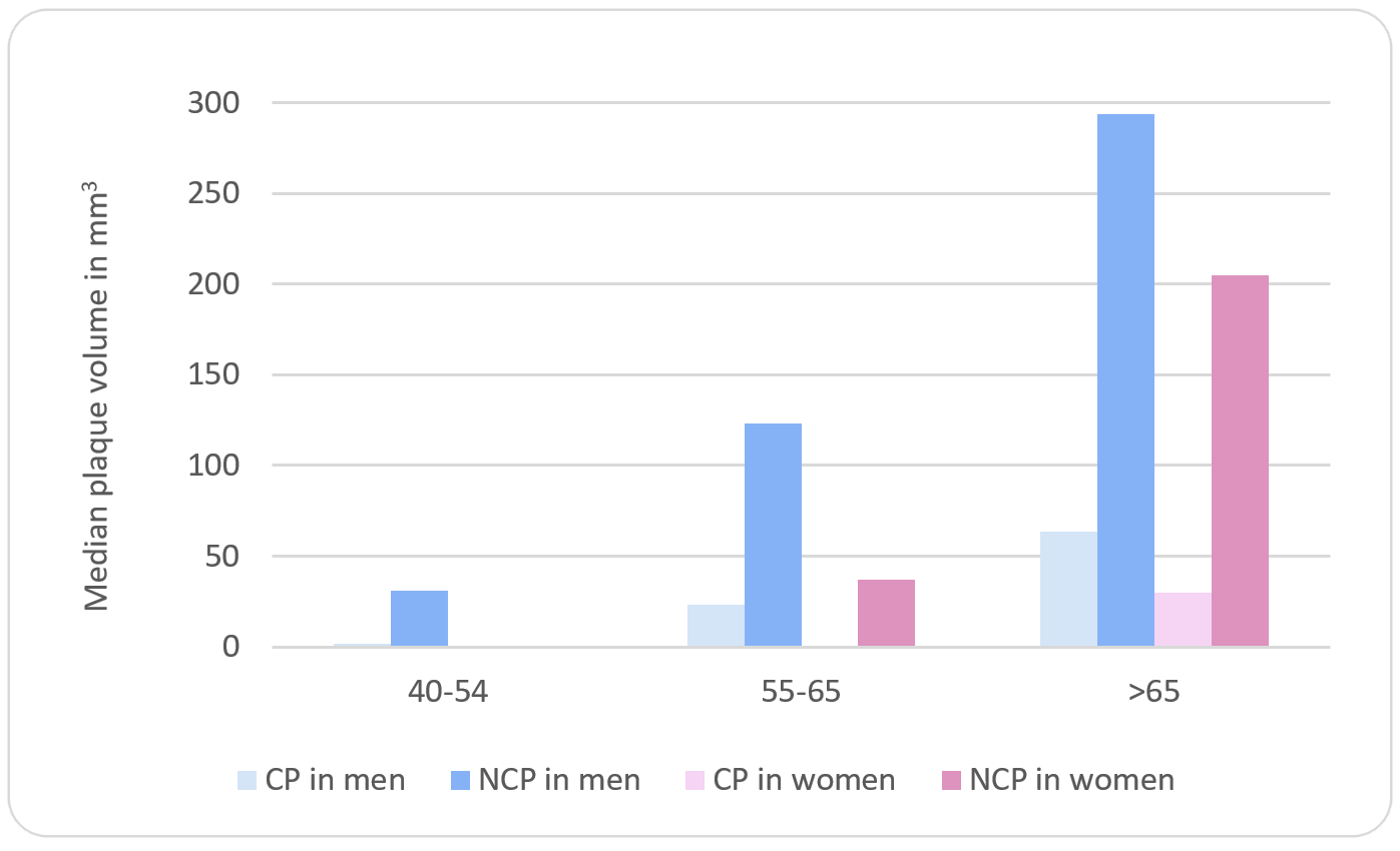
NCP is the predominant plaque subtype across all age groups, and plaque burden rises sharply after age 65 years.
CP = calcified plaque; NCP = noncalcified plaque.
Expert Opinion
In this DILWALE analysis, 63% of patients had CAC >0. In addition, men developed plaque earlier, but women showed a significant "catch-up" after the age of 65 years. This is clinically important because NCP, and particularly LAP, is a strong predictor of fatal and nonfatal myocardial infarction, regardless of CAC score. Among those with CAC ≥0, 10% of patients had NCP, a group that has lower risk overall by virtue of having small plaque volumes but may benefit from treatment to reduce long-term risk. However, prospective data from other cohorts that did not include SA adults continued to suggest a low short-term risk of an ASCVD event.4
The South Asian Woman
For SA women, reproductive risk factors and metabolic disorders such as gestational diabetes mellitus, pre-eclampsia, multiparity, premature menopause, and increased abdominal visceral adipose tissue have been strongly associated with ASCVD in prospective, observational, and registry data. The sex-stratified data are among the most clinically actionable findings in this study. Women aged >65 years demonstrated a more than fivefold increase in median total plaque compared with women who were a decade younger, rising from 44 to 255 mm3 (Figure 1). SA women aged <65 years who have a CAC ≥0 may have a low amount of NCP, and hence, treatment of such women may be reasonable to reduce long-term risk.
Thus, a potential role for AI-QCPA CTA exists in women between ages 55 and 65 years who carry multiple clinical risk factors. AI-QCPA offers prognostic value over traditional CCTA through more personalized, objective, and reproducible quantification of plaque volumes. Although relying on CAC testing can lead to a missed opportunity for earlier intervention, it is important to acknowledge that it is unknown if the extra cost and radiation exposure of CCTA would offset the potential benefit of earlier intervention. It should also be noted that AI-QCPA may overestimate plaque volumes, particularly in lower-risk patients.5
Furthermore, when there is clinical concern, earlier treatment can also be pursued without any imaging. Beyond the role of imaging, more awareness is needed regarding the prevention of ASCVD in SA adults, with emphasis on culturally tailored interventions. Screening and aggressive lifestyle modifications need to begin earlier than currently practiced in many clinical settings (Figure 2).
Figure 2: Targeted Cardiovascular Risk Assessment in South Asian Women
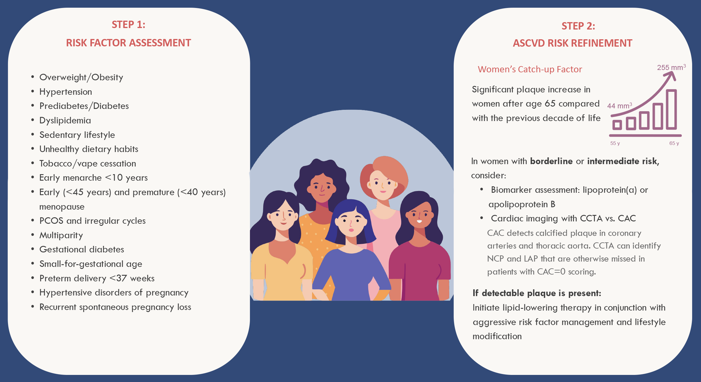
ASCVD = atherosclerotic cardiovascular disease; CAC = coronary artery calcium; CCTA = coronary computed tomographic angiography; LAP = low-attenuation plaque; NCP = noncalcified plaque; PCOS = polycystic ovary syndrome.
Conclusion
Early screening and aggressive risk factor management in SA must begin well before symptoms appear because nearly one-half of those aged 40-54 years already carry detectable plaque. Current risk scores that are often relied on may fall short, and the use of atherosclerosis imaging can have an important role when there is uncertainty about risk or treatment decisions. Although CAC testing can identify most individuals with plaque, a CAC ≥0 does not indicate a complete absence of coronary artery disease, particularly for those with intermediate or high clinical risk factors. Indeed, 10% of the study population with CAC ≥0 had NCP. Although this cohort can be classified as lower relative risk due to CAC ≥0, absolute risk remains elevated considering the presence of atherosclerosis, likely warranting targeted preventive therapy and risk factor modification.
SA women warrant distinct clinical consideration because their cardiovascular risk was underappreciated in earlier decades despite the presence of reproductive and metabolic risk-enhancing factors. Select use of AI-QCPA CTA may add diagnostic value by identifying plaque volume and subtype, particularly in women and high-risk adults. Beyond imaging, preventive efforts should focus on early and consistent identification of modifiable risk factors, namely, low-density lipoprotein cholesterol, blood pressure, blood glucose, tobacco or vape use, and overweight or obesity status.References
- Satish P, Vajramani A, Gupta S, et al. Age and sex specific coronary plaque characteristics among South Asians: Insights from the DILWALE CTA study. Am J Prev Cardiol. 2026;29:101567. Published 2026 Mar 18. doi:10.1016/j.ajpc.2026.101567
- Agarwala A, Satish P, Rifai MA, et al. Identification and management of atherosclerotic cardiovascular disease risk in South Asian populations in the U.S. JACC Adv. 2023;2(2):100258. doi:10.1016/j.jacadv.2023.100258
- Blumenthal RS, Morris PB, Gaudino M, et al. 2026 ACC/AHA/AACVPR/ABC/ACPM/ADA/AGS/APhA/ASPC/NLA/PCNA guideline on the management of dyslipidemia: a report of the American College of Cardiology/American Heart Association Joint Committee on Clinical Practice Guidelines. J Am Coll Cardiol. 2026;87(19):2624-2757. doi:10.1016/j.jacc.2025.11.016
- Sama C, Abdelhaleem A, Velu D, et al. Non-calcified plaque in asymptomatic patients with zero coronary artery calcium score: A systematic review and meta-analysis. J Cardiovasc Comput Tomogr. 2024;18(1):43-49. doi:10.1016/j.jcct.2023.10.002
- Chandrashekhar Y, Blankstein R, Shaw LJ, et al. Quantitative coronary plaque analysis in clinical practice: 2025 ACC scientific statement: a report of the American College of Cardiology. JACC Cardiovasc Imaging. 2026;19(5):637-652. doi:10.1016/j.jcmg.2025.11.008
Clinical Topics: Invasive Cardiovascular Angiography and Intervention, Noninvasive Imaging, Prevention, Interventions and Imaging, Computed Tomography, Nuclear Imaging
Keywords: Primary Prevention, Risk Assessment, Asia, Southeastern, Women, Artificial Intelligence, Computed Tomography Angiography, Asian Americans