
Pullback Pressure Gradient: Unlocking the Functional Map of CAD
Quick Takes
- Pullback pressure gradient (PPG) transforms coronary physiology from a binary ischemia test into a functional map of disease distribution.
- PPG distinguishes focal from diffuse coronary artery disease, helping identify lesions most likely to benefit from PCI.
- By enabling precision revascularization, PPG may reduce unnecessary stenting and improve PCI decision-making.
Coronary angiography has long served as the anatomical cornerstone for guiding PCI. Yet, the visual severity of a stenosis does not always reflect its physiological significance. Over the past two decades, coronary physiology, anchored by fractional flow reserve (FFR) and nonhyperemic pressure ratios (NHPRs), has transformed revascularization strategies. However, a single distal pressure value provides only a binary answer: ischemic or not. Pullback pressure gradient (PPG) assessment goes a step further, offering a dynamic and spatially resolved functional map of coronary artery disease (CAD). By tracing the distribution of pressure changes along the vessel, clinicians can differentiate focal from diffuse disease, identify target segments for intervention, and avoid unnecessary stenting.1
Findings from landmark trials such as FAME (Fractional Flow Reserve Versus Angiography for Multivessel Evaluation) and FAME 2 (Fractional Flow Reserve Versus Angiography for Multivessel Evaluation 2) established the benefit of FFR-guided PCI. Long-term pooled outcome data indicated that revascularization strategies guided by instantaneous wave-free ratio (iFR [Philips]) were associated with increased rates of 5-year major cardiovascular events compared with FFR guidance.2 However, these strategies relied on single-point measurements, summarizing total epicardial resistance into one number.
The introduction of pullback assessment overcame this limitation. By slowly withdrawing the pressure sensor from the distal to the proximal segments, during hyperemia for FFR or at rest for NHPR, operators can visualize if and where the pressure drops occur. This technique enables precise PCI planning only for lesions with a significant hemodynamic impact and avoids treatment of vessels with diffuse disease, in which the continuous progressive decrease of pressure does not identify a target for PCI. In recent years, the concept has been standardized through the PPG index, which quantifies the distribution of pressure loss along a vessel rather than relying on visual interpretation of pullback curves.
Contemporary work has refined the physiological basis of pullback measurements. PPG provides an objective metric to distinguish focal from diffuse CAD patterns, improving reproducibility compared with subjective pullback interpretation.1,3 The PPG index numerically characterizes disease distribution along a vessel, with higher values reflecting focal stenosis and lower values indicating diffuse resistance. There is a paradigm shift in physiology-guided revascularization, emphasizing PPG's role in redefining how clinicians conceptualize epicardial disease: from isolated lesions to longitudinal resistance profiles.4 This shift moves beyond threshold-based decision-making toward a pattern-based physiology. In effect, no precise cutoff has been established for PPG, instead reflecting a continuum of values moving between the two presentations of CAD. Notably, expert consensus has recently indicated values from 0.6 to 1 as certainly pointing to a focal disease whereas those <0.4 clearly reflect a diffuse pattern, thus leaving room for a large gray zone (Figure 1).
Figure 1: PPG Index for Physiological Pattern Recognition and Clinical Decision-Making
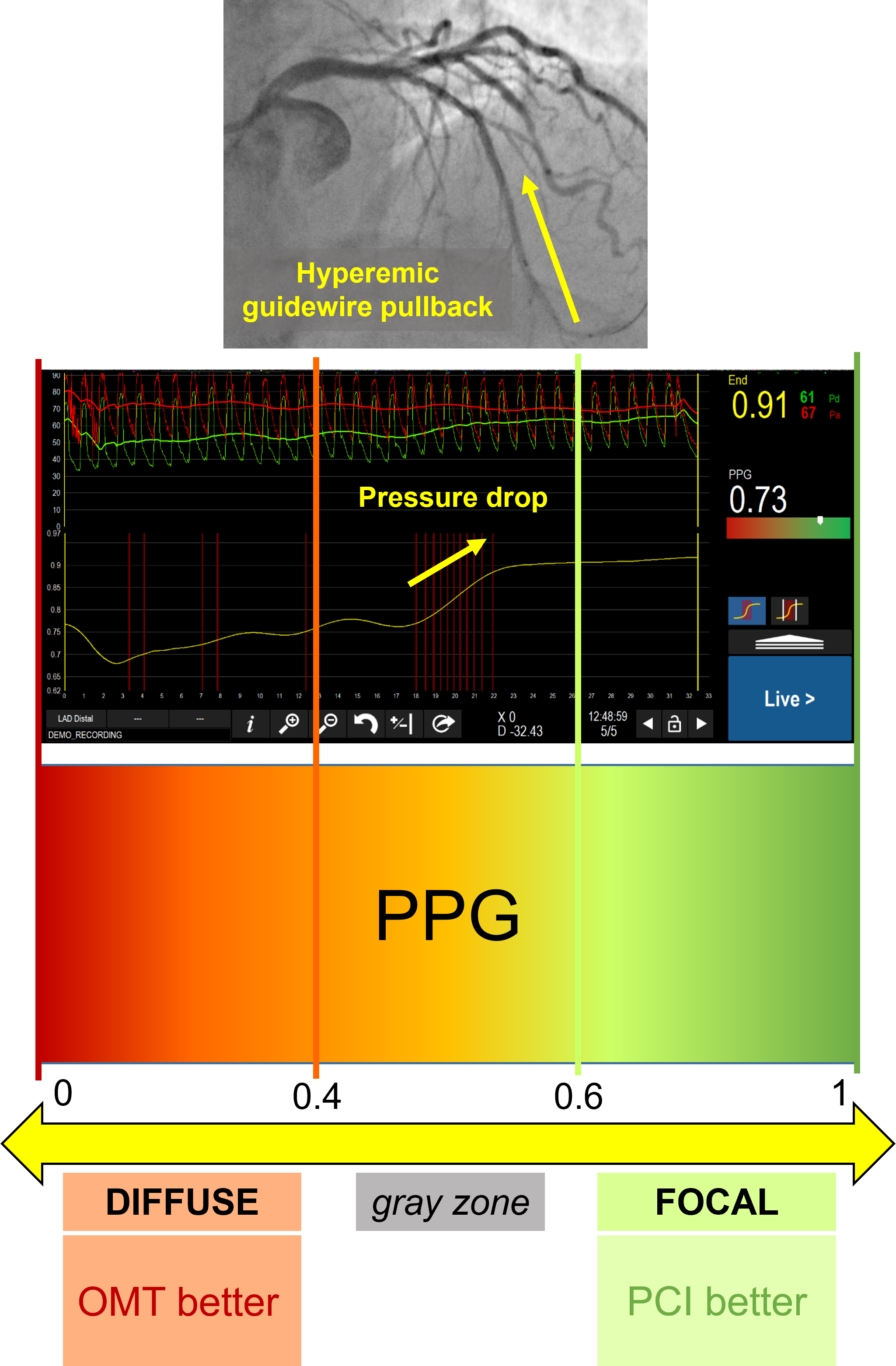
OMT = optimal medical therapy; PPG = pullback pressure gradient.
Under maximal hyperemia, microvascular resistance is minimized, isolating epicardial stenosis as the dominant determinant of pressure loss. A focal stenosis generates an abrupt step-up in pressure gradient over a short segment. In contrast, diffuse disease produces a gradual, continuous decline in pressure without discrete transitions (Figure 1). Recent mechanistic analyses confirm that focal pressure drops predict greater post-PCI physiological improvement, whereas diffuse patterns correlate with limited FFR gain after stenting.3,4 These findings reinforce the clinical importance of mapping the resistance distribution rather than treating angiographic severity alone. PPG may identify high-risk patterns even in lesions that are ischemia negative by traditional thresholds, raising the possibility that disease patterns, independently from absolute FFR values, may carry prognostic implications.5
Accurate pullback assessment begins with meticulous pressure wire preparation and equalization. After advancing the wire distally and confirming a stable baseline measurement, hyperemia is induced with adenosine. The operator then performs a slow, steady manual or motorized pullback, ideally at a uniform speed. Key technical considerations include ensuring absence of pressure drift before and after pullback, avoiding guide catheter damping, maintaining consistent hyperemia during FFR pullback. The resulting pressure trace provides a visual signature of disease distribution.
In vessels with tandem or ambiguous lesions, angiography alone may overestimate or underestimate individual lesion severity. PPG clarifies which segment contributes most to ischemia. PPG frequently alters PCI planning by identifying focal targets within otherwise diffusely diseased vessels.6 Treating only the focal, high-gradient lesion can normalize distal physiology while avoiding unnecessary stenting of noncontributory segments.
Serial stenoses interact hemodynamically, complicating interpretation of single distal measurements. PPG identifies the dominant gradient-producing lesion, guiding staged or selective intervention. After treating the primary stenosis, repeat PPG can reassess residual disease.
Diffuse disease remains challenging for PCI. When distal physiology is abnormal but no focal gradient is present, stenting long segments may yield limited improvement. Observational analyses suggest that low PPG values (diffuse pattern) predict modest post-PCI gain, supporting the optimization of medical strategies in such cases.3,4
PPG aids in understanding the branch-specific physiology in left main coronary artery or bifurcation lesions. PPG complements intravascular imaging, providing functional validation of anatomical findings.4
Emerging evidence indicates that pullback-derived metrics may offer prognostic insight beyond traditional ischemic cutoffs. Ando et al. highlighted the potential of PPG to identify hidden risk even in ischemia-negative lesions,5 whereas other findings have suggested that PPG may correlate with post-PCI physiological success and clinical outcomes.7 The integration of physiology with intravascular imaging represents a complementary strategy. Whereas imaging characterizes plaque morphology and stent optimization, pullback physiology defines the functional footprint of disease. Together, they enable precision PCI minimizing unnecessary stent length while targeting physiologically dominant segments. Despite its potential, PPG has limitations. Pressure drift, inadequate hyperemia, microvascular dysfunction, and valvular disease may affect accuracy. Moreover, most evidence is observational, and the prognostic significance of high PPG values in ischemia-negative lesions remains to be established.
Findings from recent studies position PPG as a shift from lesion-based to vessel-level physiology.4 Ongoing research is evaluating its impact on PCI outcomes, and artificial intelligence may improve reproducibility and adoption. PPG may ultimately become a routine component of physiology-guided PCI. In conclusion, PPG provides a functional map of CAD, distinguishing focal from diffuse disease and refining PCI strategy. By moving beyond binary ischemia thresholds to spatial disease characterization, PPG represents an important advance in precision coronary physiology.
References
- Dash D, Dash U, Ahmed N, Mody R. Pullback pressure gradients: redefining the functional landscape of coronary artery disease. Cardiovasc Revasc Med. Published online June 14, 2026. doi:10.1016/j.carrev.2026.06.005
- Eftekhari A, Holck EN, Westra J, et al. Instantaneous wave free ratio vs. fractional flow reserve and 5-year mortality: iFR SWEDEHEART and DEFINE FLAIR. Eur Heart J. 2023;44(41):4376-4384. doi:10.1093/eurheartj/ehad582
- Carvalho PEP, Collet C, De Bruyne B, et al. The pullback pressure gradient: a physiologic index to differentiate focal from diffuse coronary artery disease. JACC Adv. 2025;4(5):101679. doi:10.1016/j.jacadv.2025.101679
- Kechichian A, Mizukami T, Malhotra G, et al. Pullback pressure gradient: a paradigm shift in physiology-guided revascularization. Cardiovasc Revasc Med. 2025;76:1-10. doi:10.1016/j.carrev.2025.04.035
- Ando H, Collet C, Sakai K, Ohashi H, Amano T. Unmasking the hidden risk: potential implication of pullback pressure gradient in ischemia-negative lesions. Cardiovasc Interv Ther. 2025;40(4):1007-1008. doi:10.1007/s12928-025-01147-0
- Sonck J, Munhoz D, Mizukami T, Tajima A, Sakai K, De Bruyne B, Collet C, et al. Impact of the pullback pressure gradient (PPG) on PCI planning and decision-making. Eur Heart J. 2024;45(Suppl 1):ehae666.1399. doi:10.1093/eurheartj/ehae666.1399
- Munhoz D, Mizukami TM, Ikeda KI, et al.; PPG Global investigator Team. Influence of pullback pressure gradient on patient-reported and clinical outcomes in coronary artery disease, Eur Heart J. 2025;46(Suppl 1):ehaf784.1762. doi:10.1093/eurheartj/ehaf784.1762
Clinical Topics: Invasive Cardiovascular Angiography and Intervention, Stable Ischemic Heart Disease
Keywords: Coronary Stenosis, Percutaneous Coronary Intervention, Coronary Disease, Physiology, Coronary Vessels
