
DECIPHER HFpEF: CMR Tissue Mapping Tracks HFpEF Mechanisms Beyond Filling Pressure
Cardiac magnetic resonance (CMR) tissue mapping captured active relaxation impairment (σ) and passive stiffness (β) in invasively characterized heart failure with preserved ejection fraction (HFpEF), even after accounting for end-diastolic pressure (EDP) and covariates including age, sex, BMI, hypertension and diabetes, according to findings published Sept. 22 in JACC: Cardiovascular Imaging.
In the prospective multicenter DECIPHER HFpEF study, Eike C. Nagel, MD, FACC, et al., enrolled 52 patients with suspected, symptomatic HFpEF and EDP ≥16 mm Hg (mean age, 69 years; 48% women), 32 age- and sex-matched control patients (mean age, 64 years; 47% women), and 18 healthy volunteers (mean age, 32 years; 44% women). Compared with the age- and sex-matched control group, the HFpEF group had a significantly higher prevalence of hypertension (75% vs. 47%; p=0.003) and diabetes (29% vs. 6%; p=0.007) and a higher BMI (30.2 vs. 27.1; p=0.010).
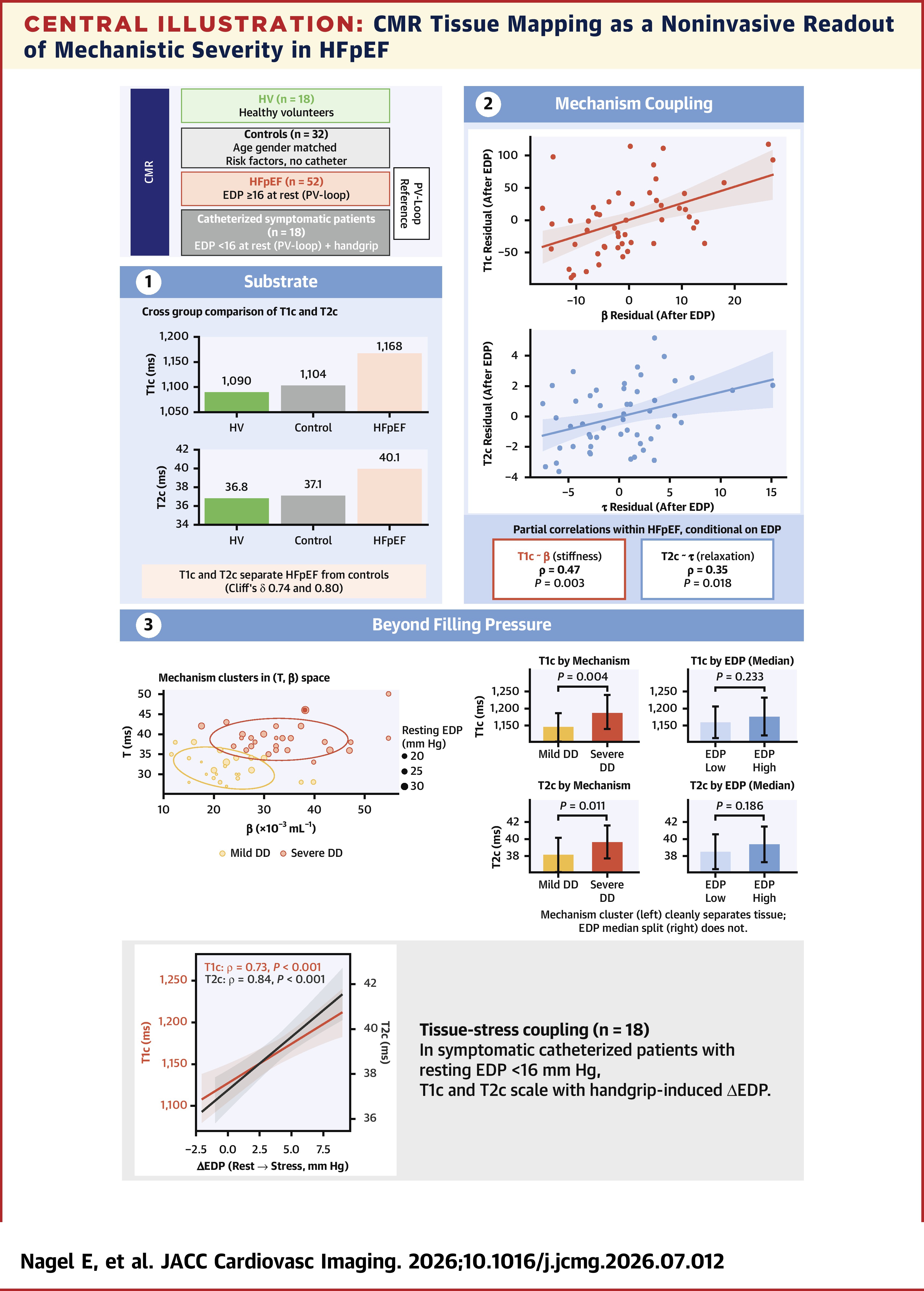
Results from CMR mapping showed that both native T1 corrected for field strength and scanner (T1c) and native T2 corrected for field strength and scanner (T2c) were higher in patients with HFpEF compared with controls (Cliff's δ, 0.74 and 0.80; p<0.001 for both). T1c correlated with β (partial ρ=0.47; p=0.003), and T2c correlated with σ (ρ=0.35; p=0.018), even after accounting for EDP. However, T1c and T2c were not associated with EDP itself.
An additional 18 catheterized patients with suspected HFpEF but EDP <16 mm Hg underwent handgrip provocation for a continuum analysis. In this subgroup, both T1c and T2c also correlated with the magnitude of handgrip-induced rise in EDP (ρ=0.73 and 0.84, respectively) and 67% of patients developed elevated filling pressures during stress despite normal resting values.
Using invasive measures of passive stiffness and active relaxation, investigators identified two distinct phenotypes within the larger HFpEF patient population: 24 patients with mild and 28 patients with severe diastolic dysfunction. These mechanism-based phenotypes were distinguished by T1c and T2c values on CMR, while stratification by EDP alone was not.
"CMR tissue mapping is therefore a noninvasive readout of mechanistic severity in HFpEF, complementary to filling pressure as a marker of disease state," write Nagel and colleagues. "The 2-phenotype mechanism-based partition observed within this cohort is hypothesis-generating; clinical application of mechanism-based stratification, and any role in informing substrate-modifying interventions, will require prospective external validation including treatment-response data. Independent cohort validation is warranted."
"The exploration of how both T1 and T2 mapping independently relate to active LV relaxation and passive chamber stiffness is a unique contribution to the literature," write Yogesh N.V. Reddy, MBBS, and Varun Sundaram, MD, on the study in an accompanying editorial comment. "These data reiterate the need to anchor noninvasive diagnostic approaches not on predicting LV diastolic dysfunction, but to focus on identification of downstream LA hypertension as the pathognomonic feature defining HFpEF."
Clinical Topics: Cardiovascular Care Team, Heart Failure and Cardiomyopathies, Acute Heart Failure
Keywords: Magnetic Resonance Spectroscopy, Ventricular Dysfunction, Left, Heart Failure, Heart Failure, Preserved Ejection Fraction