Multimodality CV Imaging in the Evaluation of Cardiac Sarcoidosis
In a State-of-the-Art Review published Oct. 6 in JACC: Cardiovascular Imaging, Katharine E. Thomas, MBBS, and colleagues examine the evolving role of multimodality cardiovascular imaging in the diagnosis and management of cardiac sarcoidosis (CS), including an appraisal of the current guideline recommendations, a review of imaging advances, a summary of ongoing gaps in understanding, and guidance for using imaging to improve diagnostic accuracy, risk assessment and treatment decisions.
CS remains substantially underdiagnosed, with approximately 5% of cases detected clinically, while autopsy studies reveal cardiac involvement in up to 30%. Symptoms may be nonspecific, and CS can mimic other cardiac conditions.
Endomyocardial biopsy can establish a definitive diagnosis but is invasive and can have a sensitivity as low as 20% due to the patchy distribution of the disease. Echocardiography can identify overt structural abnormalities but lacks sensitivity for CS and may miss cases without morphological or electrical abnormalities.
Against this backdrop, the review highlights the importance of cardiac magnetic resonance (CMR) and fluorodeoxyglucose (FDG)-PET, both of which have a Class I indication for diagnostic evaluation of CS in the 2025 European Society of Cardiology Myopericarditis guidelines. Cardiac symptoms, rhythm or structural abnormalities, or elevated cardiac biomarkers should prompt evaluation for CS. CMR can assess cardiac structure and function, fibrosis with late gadolinium enhancement (LGE) and inflammation or edema using T2-weighted imaging and T1/T2 mapping. FDG-PET provides complementary assessment of active inflammation and should be paired with perfusion imaging.
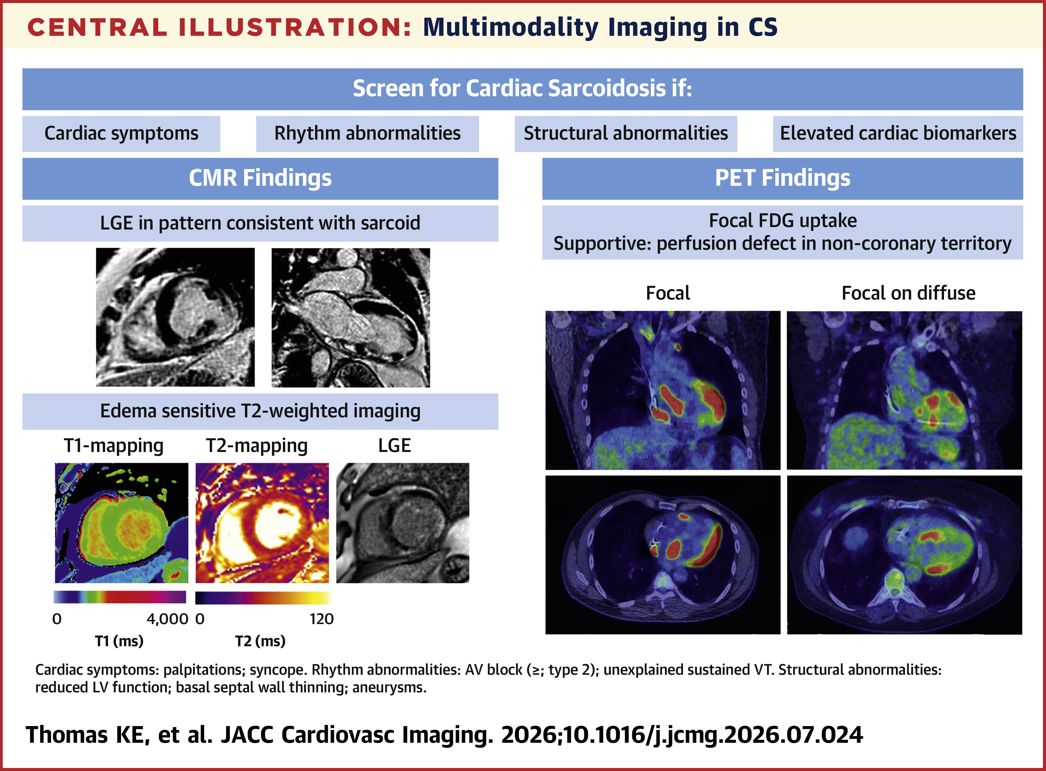
The review also illustrates the heterogeneity of CS and the complementary strengths of different modalities. CMR can demonstrate a range of LGE patterns, while T1/T2 mapping may identify myocardial inflammation even without LGE. PET can demonstrate different patterns of FDG uptake, while delayed enhancement CT may provide information about myocardial fibrosis when CMR is unsuitable, although further validation is needed.
Important uncertainties remain, including who to screen, how to interpret discordant CMR and PET results, and how imaging should guide treatment. There is no consensus on what level of LGE burden is clinically significant, no standardized uptake value (SUV) threshold on PET to distinguish CS from normal myocardium, and no consensus on what reduction in SUV represents an acceptable treatment response. Evidence for T1/T2 mapping remains largely limited to smaller, single-center studies, while hybrid PET/CMR is constrained by availability, cost and technical challenges.
Nevertheless, "advanced imaging modalities, such as CMR, should be offered more routinely, especially for patients with symptoms and signs suspicious for CS," Thomas and colleagues ultimately recommend. "Advanced imaging protocols should be designed to image both fibrosis and inflammation while providing comprehensive coverage of the heart (including the [right ventricle]), given that CS can be a patchy disease."
Clinical Topics: Heart Failure and Cardiomyopathies, Noninvasive Imaging, Computed Tomography, Nuclear Imaging
Keywords: Magnetic Resonance Spectroscopy, Fluorodeoxyglucose F18, Fibrosis, Tomography, X-Ray Computed, Perfusion Imaging, Sarcoidosis, Inflammation