A 56-year-old man with a history of obesity, type II diabetes mellitus, chronic obstructive pulmonary disease, hyperlipidemia and coronary artery disease and recent cavo-tricuspid isthmus-dependent atrial flutter ablation presents with palpitations and increased dyspnea.
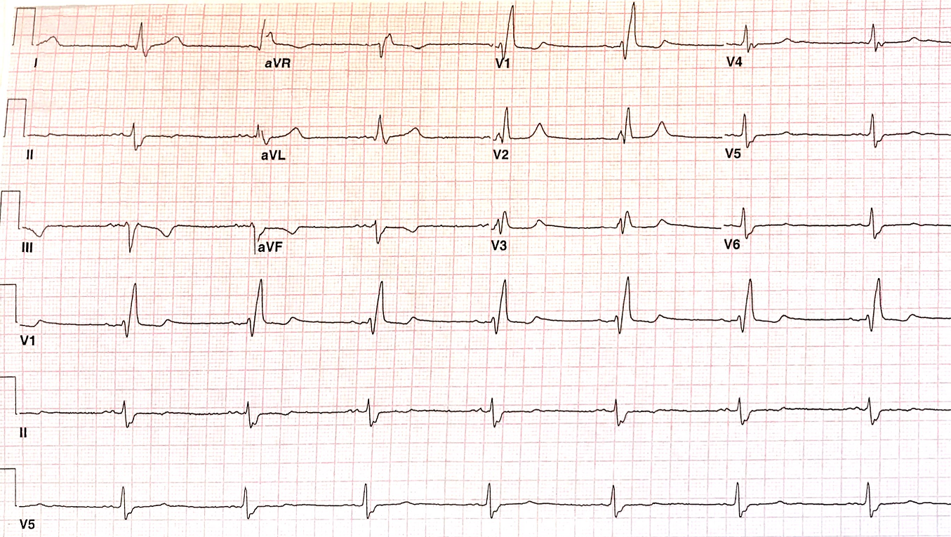
Figure 1
Figure 1
The ECG is suggestive of which of the following:
Show Answer
The correct answer is: E. All of the above.
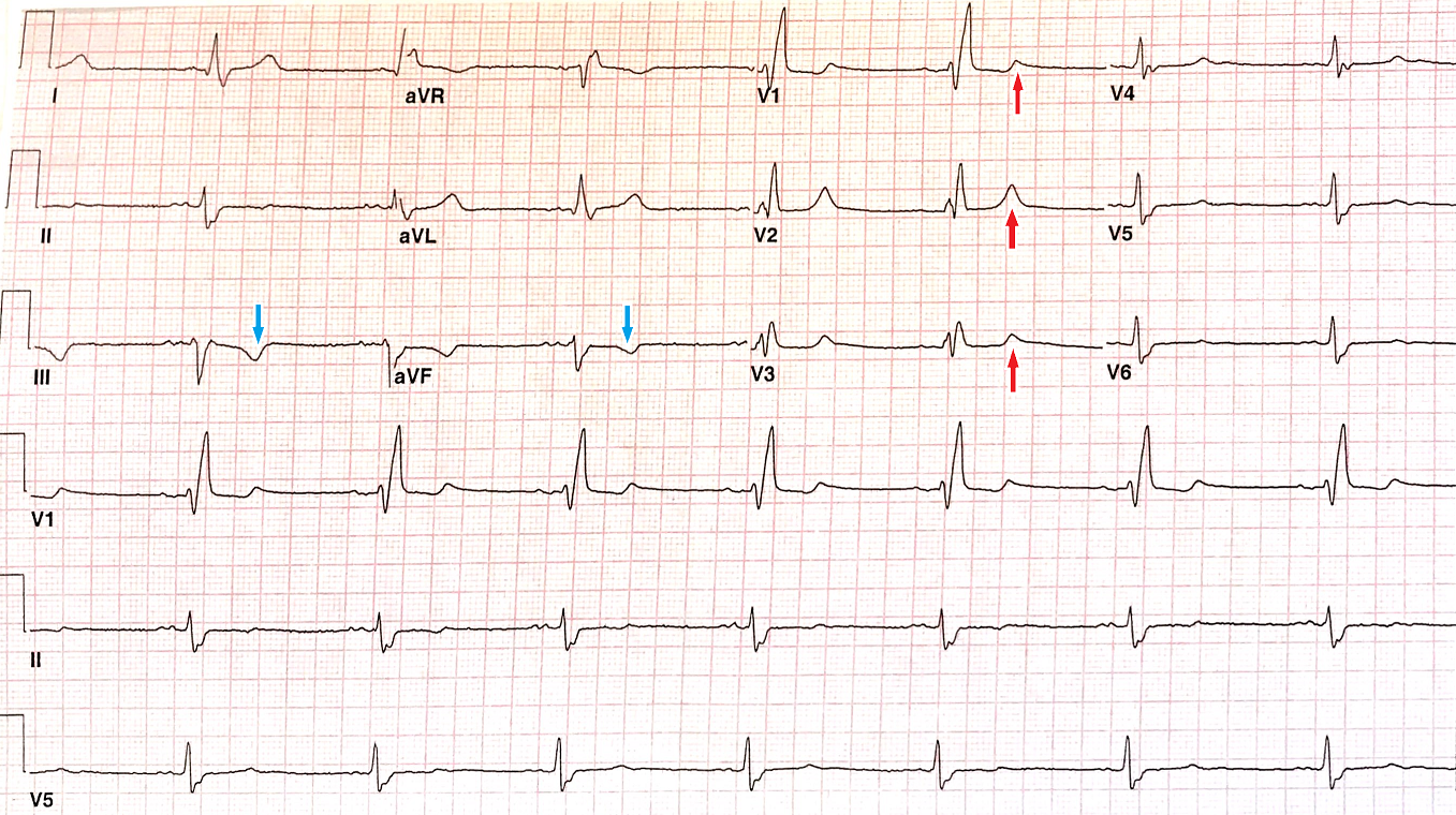
The patient has sinus rhythm with P wave of prolonged duration. The axis is leftward (approximately 40 degrees) with a QRS typical for right bundle branch block (RBBB). The T wave is upright in V1 through V3, which is not typical for RBBB, indicative of posterior ischemia (red arrows); concomitantly, the T waves are inverted in lead III and aVF indicating inferior wall involvement (blue arrows) as well.
Figure 2
Figure 2
Abnormal inferior and postero-lateral wall motions noted on cardiac imaging.
Radiofrequency ablation of the cavo-tricuspid isthmus dependent atrial flutter could result in right coronary artery injury resulting myocardial injury/ischemia especially with medial/septal approach of the ablation procedure.1,2
References
Pothineni NV, Kancharla K, Katoor AJ, et al. Coronary artery injury related to catheter ablation of cardiac arrhythmias: a systematic review. J Cardiovasc Electrophysiol 2019;30:92-101.
Al Aloul B, Sigurdsson G, Can I, Li JM, Dykoski R, Tholakanahalli VN. Proximity of right coronary artery to cavotricispid isthmus as determined by computed tomography. Pacing Clin Electrophysiol 2010;33:1319-23.