
Recurrent Myocarditis in a Child: More Than Meets the MRI
A 10-year-old boy was admitted to the hospital for management of acute chest pain and troponin elevation with peak level 54 ng/mL (reference range <0.03 ng/mL). An electrocardiogram had findings of sinus rhythm, right-axis deviation, and nonspecific ST and T-wave abnormalities. Cardiac magnetic resonance imaging (cMRI) had findings of normal left ventricular (LV) ejection fraction (EF) and right ventricular (RV) EF (LV, 59%; RV, 51%). Tissue characterization sequence findings were notable for subepicardial late gadolinium enhancement (LGE) in the basal inferolateral LV segment with associated myocardial edema, meeting modified Lake Louise Criteria for acute myocarditis (Figure 1). An infectious workup had unrevealing findings. He was discharged from the hospital with resolution of chest pain and downtrending troponin levels.
Figure 1: SAX View on cMRI Showing Elevated Relaxation Times and LGE Within the Basal Inferolateral LV Segment
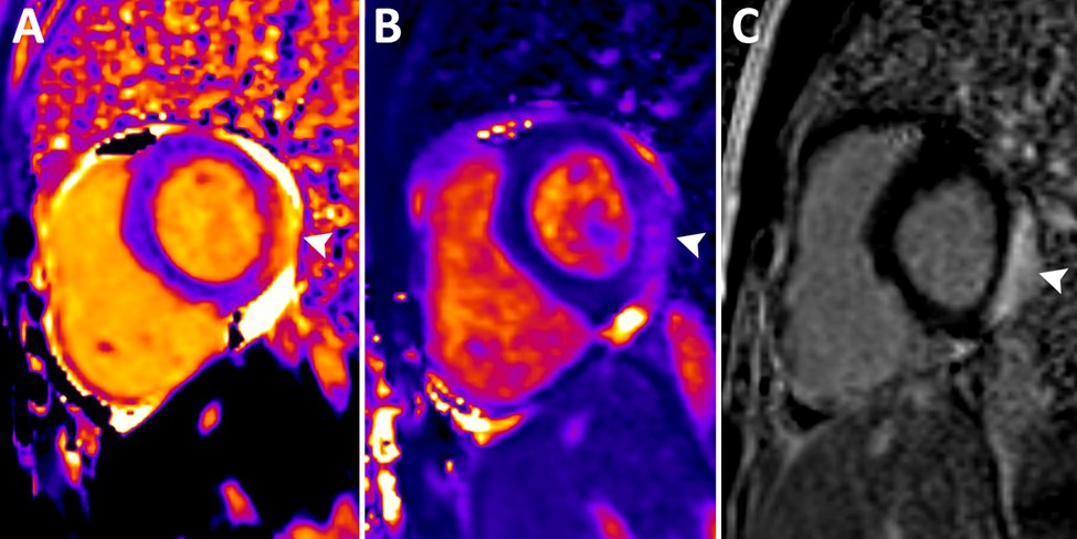
cMRI showing elevated relaxation times within the basal inferolateral LV segment on precontrast T1 mapping (panel A) and T2 mapping (panel B) sequences in the SAX view. (Panel C) Subepicardial LGE of the basal inferolateral LV segment. White arrows indicate the locations of the specified pathology in each panel.
cMRI = cardiac magnetic resonance imaging; LGE = late gadolinium enhancement; LV = left ventricular; SAX = short-axis.
He presents 1 year later with a recurrence of chest pain and troponin level elevation. A repeat cMRI has findings of mildly depressed LV systolic function (EF 51%), mildly depressed RV systolic function (EF 43%), dyskinesis of the inferior RV wall, new subendocardial LGE of the inferior and septal walls of the RV, and evidence of fatty infiltration of the LV lateral wall (Figure 2). Rhythm monitoring reveals brief runs of accelerated idioventricular rhythm.
Figure 2: cMRI Cine Steady-State Free Precession Imaging of the Mid and Apical LV lateral Wall and Subendocardial LGE of the RV Inferior and Septal Walls
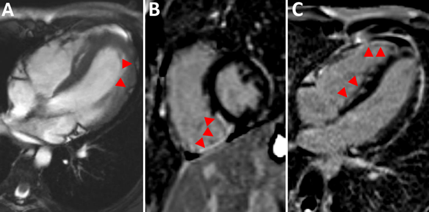
(Panel A) cMRI cine steady-state free precession imaging of macroscopic fatty infiltration of the mid and apical LV lateral wall. Subendocardial LGE of the RV inferior and septal walls in the SAX (panel B) and 4Ch (panel C) views. Red arrows indicate the locations of the specified pathology in each panel.
4Ch = four-chamber; cMRI = cardiac magnetic resonance imaging; LGE = late gadolinium enhancement; LV = left ventricular; RV = right ventricular; SAX = short-axis.
Which one of the following is the most appropriate next step in his management?
Show Answer