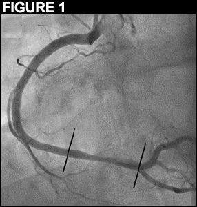
A 32-year-old male presents to the emergency department with stuttering chest discomfort. ECG confirms transient ST elevations in the inferior leads, which rapidly subside in the following minutes. By coronary angiography we find a right dominant coronary system and single vessel coronary artery disease with haziness in the distal segment of the right coronary artery just before the crux and accompanying TIMI 3 flow (Figure 1; Video 1).
What would you recommend next?
Show Answer
The correct answer is: 4. Proceed with intravascular imaging
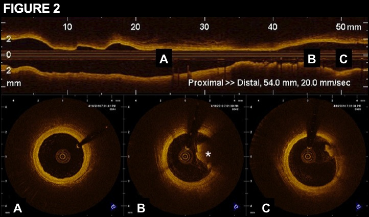
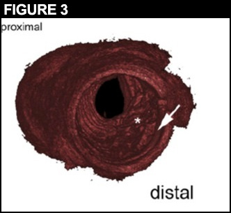
We proceeded with intravascular imaging to get a more accurate appreciation of the pathophysiology at hand given the clinical consequences for this young patient. We performed optical coherence tomography (OCT) to scan the region of interest (Video 2). We demonstrated significant intimal thickening and a significant load of mural, predominantly white thrombus in a region of focal, eccentric lipid-rich plaque formation. OCT is an invasive imaging tool enabling endoluminal scanning of coronary arteries with high resolution and in 3D bearing in mind that the role for OCT in routine clinical decision-making has not been formally established (Figures 2 and 3)(1). Subsequently we performed thrombectomy, added intracoronary Glycoprotein 2b3a antagonists and treated the diseased segment with a drug eluting stent.
References
Levine GN, Bates ER, Blankenship JC, Bailey SR, Bittl JA, Cercek B, et al. 2011 ACCF/AHA/SCAI Guideline for Percutaneous Coronary Intervention. A report of the American College of Cardiology Foundation/American Heart Association Task Force on Practice Guidelines and the Society for Cardiovascular Angiography and Interventions. J Am Coll Cardiol 2011; 58:e44-122.