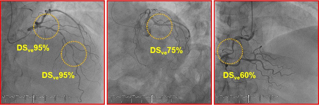
A 70-year-old man with a history of hypertension, hypercholesterolemia, and smoking started complaining about chest pain with mild exertion (Canadian Cardiovascular Society Class III) and thus consulted a cardiologist. A stress electrocardiography test was performed showing symptoms and 1.5 mm ST depression in V4-V5-V6 at the peak of the exercise. Furthermore, a computed tomography (CT) scan was performed showing 3-vessel coronary disease, with a calcium score over 400. Subsequently, the patient underwent coronary angiography that confirmed the results of the CT scan, showing two long, tight lesions in the context of a diffusely diseased left anterior descending (LAD) artery, a 75% lesion at the ostium of a large marginal branch, and a 60% stenosis of the mid right coronary artery (RCA) (Figure 1). The calculated SYNTAX score was 25.
Figure 1
Which of the following statements is correct?
Show Answer
The correct answer is: C. Physiological assessment with FFR might potentially downgrade the anatomic severity of coronary disease.
American and European revascularization guidelines state that in patients with 3-vessel disease and a SYNTAX score ≥22, surgical treatment with coronary artery bypass grafting is preferable over PCI. In particular, European guidelines report a Class III contraindication to treat such a patient with PCI.1,2
On the other hand, studies in the past have shown that it is beneficial to revascularize a stenosis only if myocardial ischemia is relevant, otherwise medical therapy is preferable.3 As opposed to noninvasive stress tests (i.e., SPECT) that might result in false negative results in case of 3-vessel disease, FFR is independent from both blood flow and the status of microcirculation in myocardial territories supplied by a different coronary artery. In fact, FFR has an unsurpassed spatial resolution, being able to detect ischemia-causing lesions at a segment level. FFR measurement in stable patients with multivessel disease is reliable and represents one of the main indications of FFR since the FAME (Fractional Flow Reserve versus Angiography for Guiding Percutaneous Coronary Intervention) trial demonstrated the superiority of an FFR-guided PCI strategy over an angiography-guided one.4 Of note, surgical revascularization guided by FFR was shown not to increase cardiovascular events at 36 months compared with an angiography-guided strategy and to simplify the surgical protocol.5
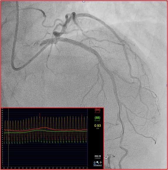
In this patient, FFR was measured in all 3 vessels, showing a non-significant value in the RCA and the marginal branch (0.92 and 0.84, respectively) and an expected very significant value in the LAD (0.40). Thus, the anatomical 3-vessel disease was reduced to a functional single-vessel, disease and the operator chose to perform PCI with implantation of two drug-eluting stents. The final angiographic result was excellent, with Thrombolysis in Myocardial Infarction 3 flow and post-PCI FFR at 0.93 (Figure 2).
Figure 2
References
Patel MR, Calhoon JH, Dehmer GJ, et al. ACC/AATS/AHA/ASE/ASNC/SCAI/SCCT/STS 2017 Appropriate Use Criteria for Coronary Revascularization in Patients With Stable Ischemic Heart Disease: A Report of the American College of Cardiology Appropriate Use Criteria Task Force, American Association for Thoracic Surgery, American Heart Association, American Society of Echocardiography, American Society of Nuclear Cardiology, Society for Cardiovascular Angiography and Interventions, Society of Cardiovascular Computed Tomography, and Society of Thoracic Surgeons. J Am Coll Cardiol 2017;69:2212-41.
Windecker S, Kolh P, Alfonso F, et al. 2014 ESC/EACTS Guidelines on myocardial revascularization: The Task Force on Myocardial Revascularization of the European Society of Cardiology (ESC) and the European Association for Cardio-Thoracic Surgery (EACTS)Developed with the special contribution of the European Association of Percutaneous Cardiovascular Interventions (EAPCI). Eur Heart J 2014;35:2541-619.
Hachamovitch R, Hayes SW, Friedman JD, Cohen I, Berman DS. Comparison of the short-term survival benefit associated with revascularization compared with medical therapy in patients with no prior coronary artery disease undergoing stress myocardial perfusion single photon emission computed tomography. Circulation 2003;107:2900-7.
Tonino PA, De Bruyne B, Pijls NH, et al. Fractional flow reserve versus angiography for guiding percutaneous coronary intervention. N Engl J Med 2009;360:213-24.
Toth G, De Bruyne B, Casselman F, et al. Fractional flow reserve-guided versus angiography-guided coronary artery bypass graft surgery. Circulation 2013;128:1405-11.