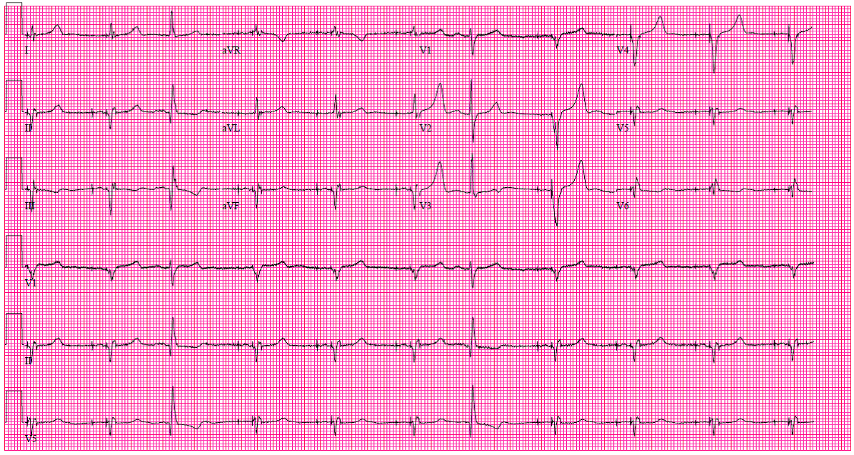
A 72-year-old man with history of coronary artery disease, hypertension, cerebrovascular accident, hyperlipidemia, and ischemic cardiomyopathy. His left ventricular ejection fraction is 35%. He is seen because of a one-week increase in dyspnea and orthopnea. An electrocardiogram (ECG) is performed (Figure 1).
Figure 1:
The ECG shows which of the following?
Show Answer
The correct answer is: E. B and D.
The ECG shows appropriate demand atrioventricular pacing. Beats 3 and 7 are premature atrial beats, which are conducted with normal atrioventricular conduction and QRS demonstrating inferior infarction of undetermined age with prolonged QRS duration. The initial and terminal forces are approximately 180 degrees, which represents infarction block or intraventricular conduction defect (IVCD).
Intraventricular conduction defect (IVCD) following myocardial infarction is common. The QRS is usually wide (> or equal to100 milliseconds: "it is approximately 130 milliseconds in our patient"). The QRS forces point leftward and superiorly producing Q waves in leads II, III, and aVF with slow terminal forces directed 180 degrees opposite the inferior Q's, producing an S wave in lead aVL and lead I, which is not seen well in this ECG. This is indicative of infarction block of the inferior wall. Though the changes in the QRS have been historically labeled as infarction block in patients with myocardial infarction, they are not specific and have been described in patients with non-ischemic cardiomyopathy, myocardial fibrosis, and chronic lung disease with emphysema, and in patients without lung or heart disease. Therefore, intraventricular conduction defect (IVCD) has been used instead of infarction block.
The R > S in V2 with upright T wave in V1 suggests posterior infarction as well.
References
C. Hilmon Castle, William M Keane. Electrocardiographic "Peri-Infarction Block" A Clinical and Pathologic Correlation. Circulation 1965;31:403-8.
Gruber JS, Stair B, Aktas M, Bravo-Jaimes K. Left Bundle Branch Block and Complete Heart Block Complicating Inferior Myocardial Infarction. Ann Noninvasive Electrocardiol 2017 Jan;22.