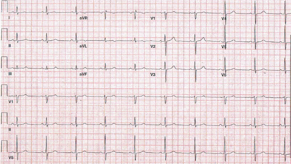
A 60-year-old obese man presents with a history of hypertension, hyperlipidemia, obstructive sleep apnea, diabetes mellitus type II and gastroesophageal reflux disease (GERD). He presents with an increasing shortness of breath and anterior chest discomfort. An electrocardiogram (ECG) is performed (Figure 1).
Figure 1
The correct answer is: A. Suggestive of postero-lateral ischemia.
The QRS–T wave angle is wide in both horizontal and frontal planes, with T wave positive in V1 and inverted in aVL with QRS axis of + 45 degrees. The T wave is biphasic in lead I.
The patient had high grade obstruction in a diagonal branch with 50% obstruction in stent of the right coronary artery (RCA).
References
- Andersen MP, Terkelsen CJ, Sørensen JT, et al. The ST injury vector: electrocardiogram-based estimation of location and extent of myocardial ischemia. J Electrocardiol 2010;43:121-31.
- Schwaab B, Katalinic A, Riedel J, Sheikhzadeh A. Pre-hospital diagnosis of myocardial ischaemia by telecardiology: safety and efficacy of a 12-lead electrocardiogram, recorded and transmitted by the patient. J Telemed Telecare 2005;11:41-4.

