A 46-year-old male patient with a past medical history of Hodgkin's stage IIb lymphoma at age 13 treated with chemotherapy (agents unknown) and radiation therapy presents for worsening shortness of breath over the past 20+ years, though it was been notably worse over the past couple months. For most of his adult life, he has felt like he could not exert himself like his peers. Now he can barely climb a flight of stairs. Videos 1-2 and Figures 1-4 show the echocardiography and catheterization findings.
Video 1
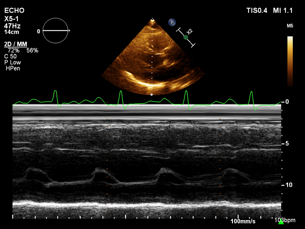
Figure 1
Figure 1
Video 2
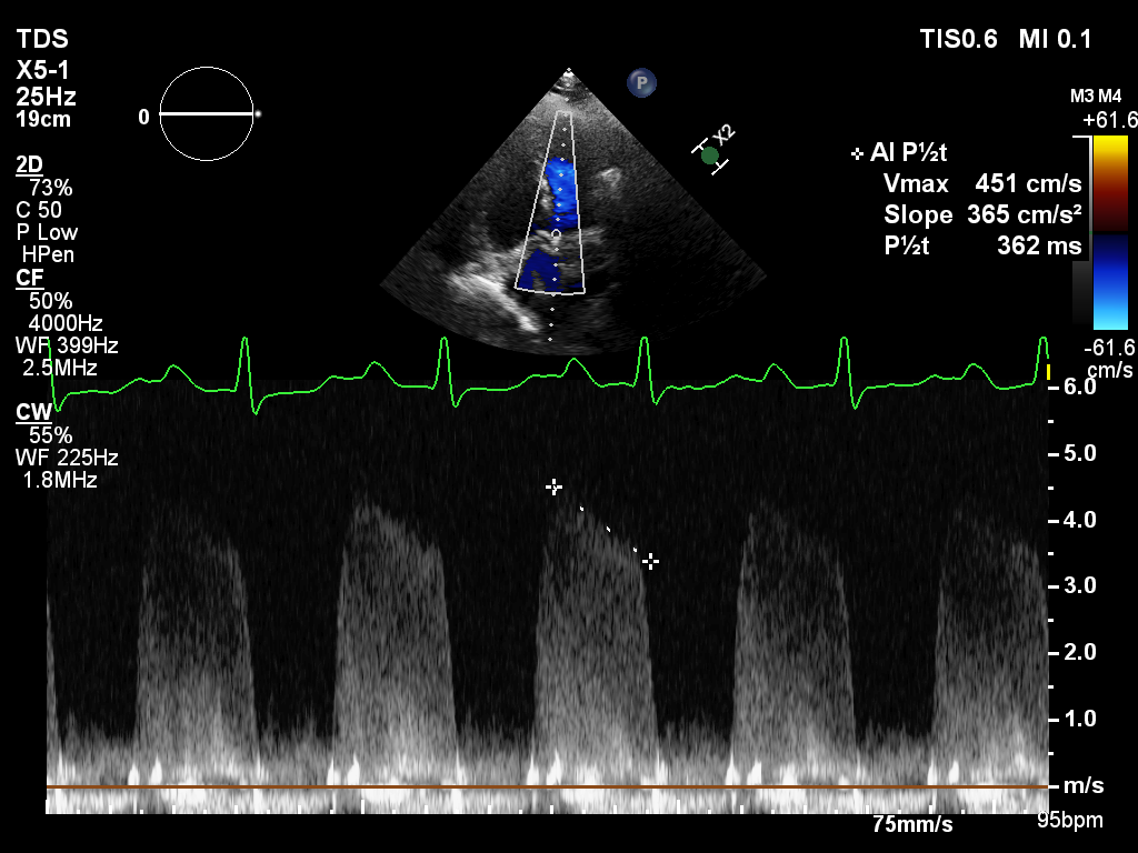
Figure 2
Figure 2
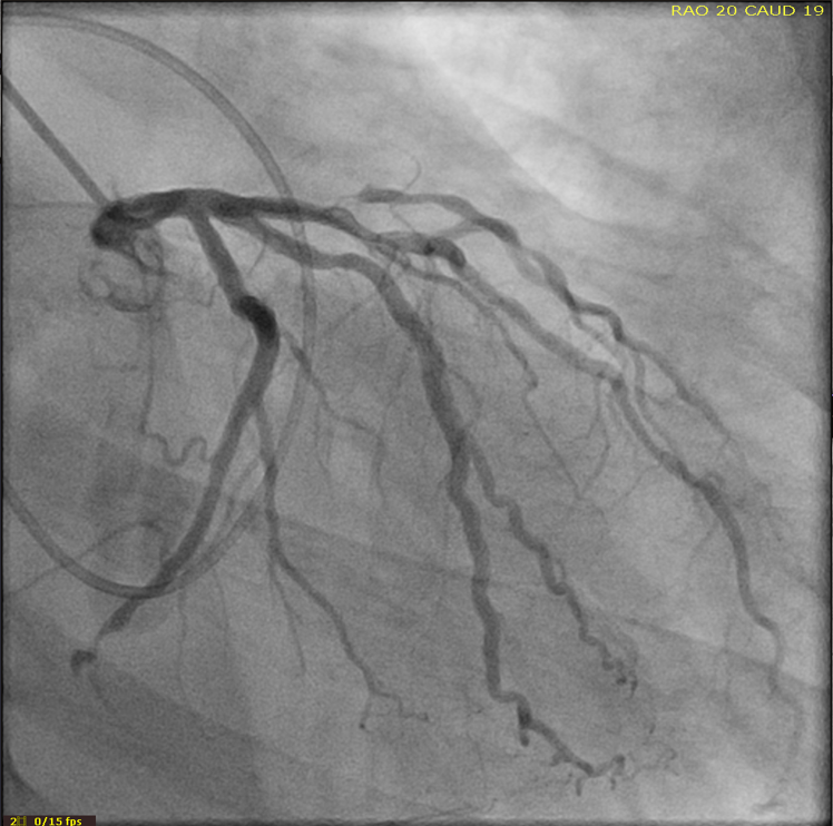
Figure 3
Figure 3
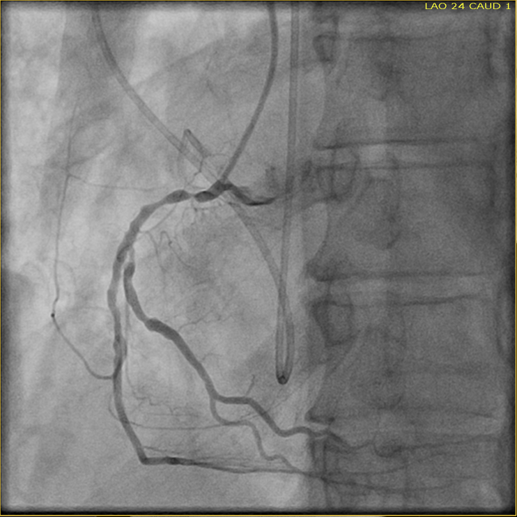
Figure 4
Figure 4
Based on the echocardiography and catheterization findings, what is the primary etiology of his heart disease?
Show Answer
The correct answer is: D. Radiation-induced heart disease
This patient has radiation-induced heart disease causing multiple findings, including mixed mitral (Video 2) and aortic valve disease (Figure 2) and severe coronary artery disease (Figures 3-4) with decreased ejection fraction at 40%. The right coronary cusp of the aortic valve is fixed (Video 1). The mitral posterior leaflet is fixed (Figure 1). The aorto-mitral curtain is thick and calcified. A subsequent catheterization showed significant coronary disease in the proximal diagonal, distal left circumflex, and proximal right coronary artery. All of these lesions are well within the field of prior mantle field radiation with conventional beam therapy, which was used extensively with high cardiac doses from the 1960s to the 1990s for treatment of Hodgkin's lymphoma.
References
Cutter DJ, Schaapveld M, Darby SC, et al. Risk of valvular heart disease after treatment for Hodgkin lymphoma. J Natl Cancer Inst 2015;107:djv008.
van Nimwegen FA, Schaapveld M, Cutter DJ, et al. Radiation Dose-Response Relationship for Risk of Coronary Heart Disease in Survivors of Hodgkin Lymphoma. J Clin Oncol 2016;34:235-43.
Lancellotti P, Nkomo VT, Badano LP, et al. Expert consensus for multi-modality imaging evaluation of cardiovascular complications of radiotherapy in adults: a report from the European Association of Cardiovascular Imaging and the American Society of Echocardiography. J Am Soc Echocardiogr 2013;26:1013-32.