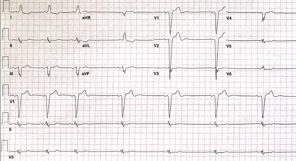
A 75-year-old man with recent therapy for esophageal squamous cell carcinoma presents to the the emergency department with palpitations, weakness, and dysphagia. He is initially in atrial fibrillation, and has history of hypertension. The following ECG is obtained (Figure 1).
Figure 1
Figure 1
The ECG shows which of the following?
Show Answer
The correct answer is: E. Blocked atrial premature depolarization.
The ECG shows sinus rhythm with left bundle branch block (LBBB). The P waves are approximately 140 milliseconds in duration indicating diffuse intra-atrial disease. The axis is borderline leftward (-28 degree) with poor R wave progression in the precordial leads, which is typical for LBBB with left axis deviation. An old anterior infarct cannot be excluded. The ST-T vectors are typical for LBBB and not indicative of ischemia.
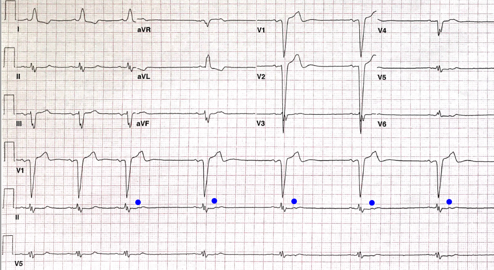
The rhythm shows 3 beats of sinus rhythm at approximately 60 beat per minutes (bpm) followed by regular sinus beats at approximately 40 bpm. Inspection of the T wave prior to each bradycardic beat shows a non-conducted atrial premature depolarization (blue dots in Figure 2).
Figure 2
Figure 2
These very atrial premature depolarization (APD) occur when the AV node is in absolute refractory period. These blocked atrial bigeminy will appear similarly to sinus bradycardia — close inspection for these subtle APDs is imperative to avoid misdiagnosis and unnecessary testing.
References
Akdeniz C, Tanidir IC and Tuzcu V. Blocked atrial bigeminy presenting with bradycardia. Congenit Heart Dis 2012;7:E82-4.