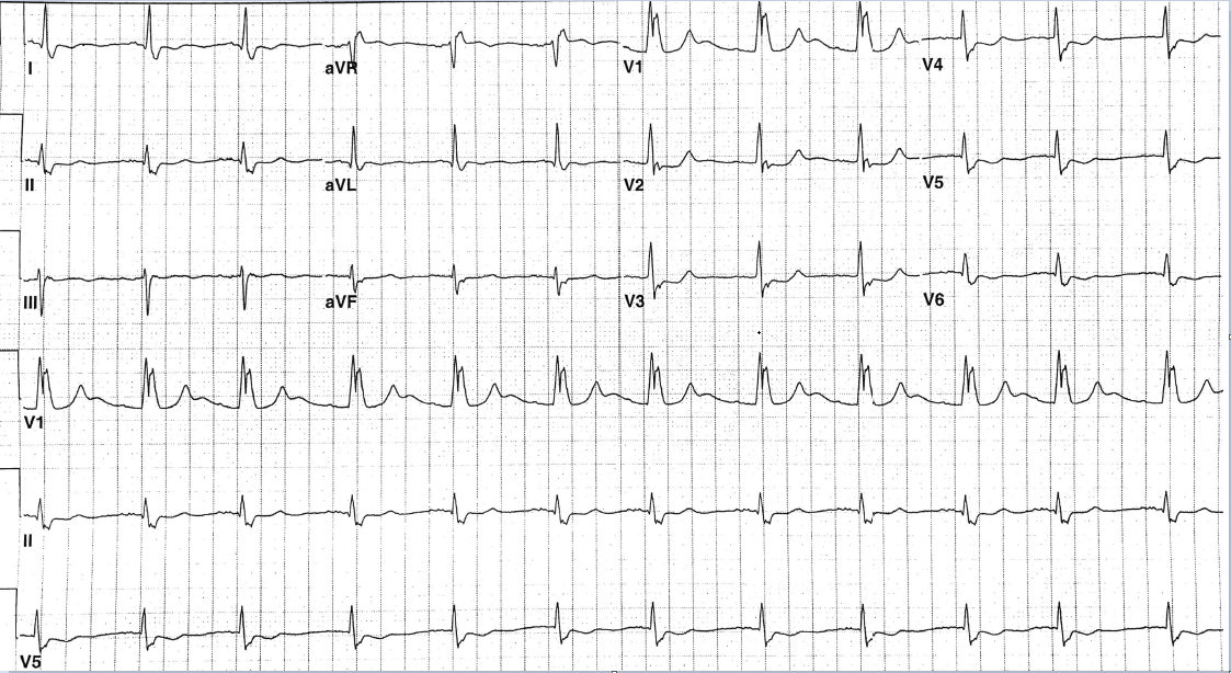
The patient is a 77-year-old man with a history of type 2 diabetes mellitus, coronary artery disease s/p 3v CABG, and bioprosthetic AVR 20 years ago, paroxysmal atrial fibrillation on warfarin, and angioectasia. He is seen in emergency department because of abdominal pain, melena and chest heaviness for 3 days. His INR was found to be 13, hemoglobin 8.7 and troponin 1.10. An ECG was performed (Figure 1):
Figure 1
The correct answer is: D. Recent postero-lateral infarction.
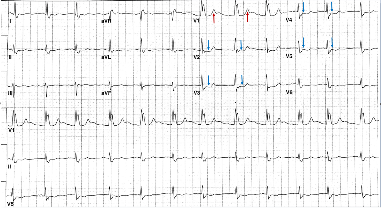
The ECG shows sinus rhythm at a rate of 66 beats/minute. The QRS axis is approximately -20 degree. The QRS is wide secondary to right bundle branch block. The R wave in V1 and V2 is prominent (R>S). Additionally, The T wave is upright in V1 (red arrow) and the ST depression in V2, V3, V4, V5 (blue arrow) as reflection of recent postero-lateral infarction.
Figure 2
References
- Cornejo-Guerra JA, Manzur-Sandoval D, Guadalajara-Boo JF, Briseno-de la Cruz JL. Case report: posterior myocardial infarction in presence of right bundle branch block: an old concept with new findings. Eur Heart J Case Rep 2018;2.


