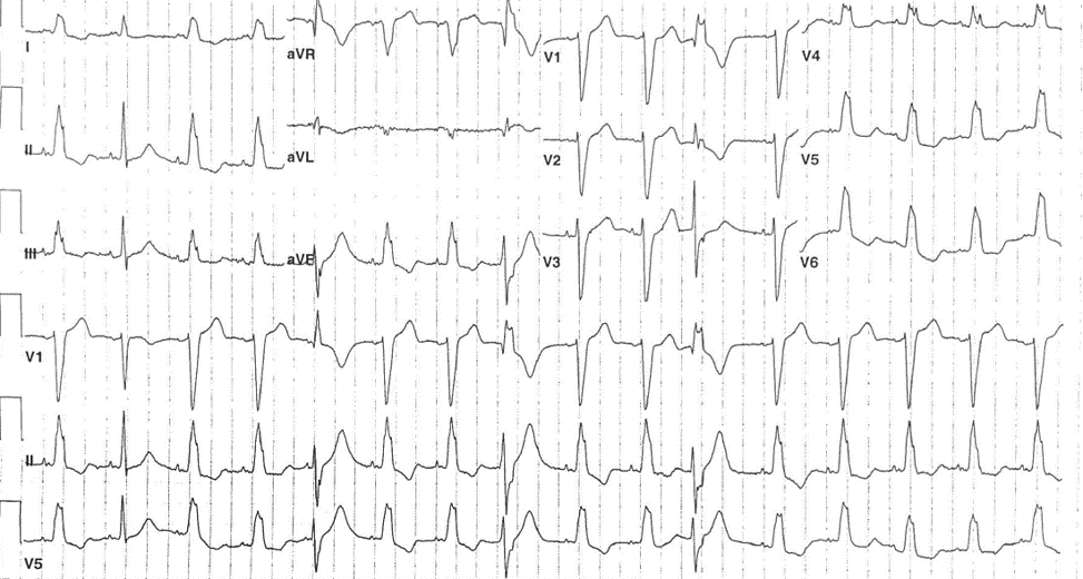
A 61-year-old woman with a history of hypertension, chronic hepatitis B, coronary artery disease, and pancreatic disease with cysts presents to the emergency department with abdominal pain, nausea, and vomiting. The following ECG is performed:
The correct answer is: D. Junctional beats with aberration.
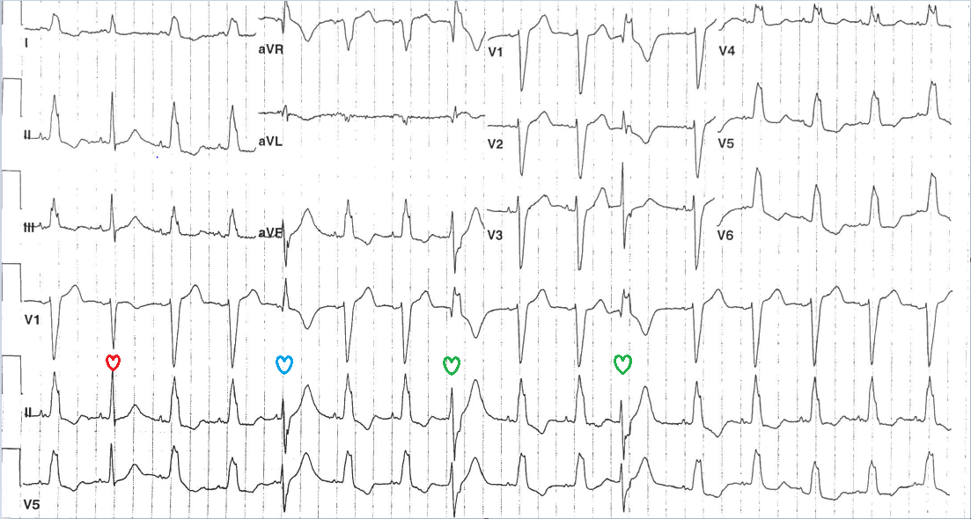
The ECG shows sinus rhythm with abnormal P wave with a QRS of left bundle branch block (LBBB) morphology. The ST- T wave abnormalities are typical for LBBB. The second, fifth, eight, and 11th beats show initially a narrow QRS which could indicate equal conduction over both bundle (second beat/red heart) followed by incomplete RBBB and left axis deviation (LAD) (fifth beat/blue heart) then complete RBBB with LAD (eighth and 11th beats/green hearts). This likely represents a junctional focus with different degree of aberration. The shorter the interval between the QRS complex of sinus and junctional beat, the higher the degree of aberrancy.
References
- Sandler IA, Marriot H. The differential morphology of anomalous ventricular complexes of RBBB-type in lead V; ventricular ectopy versus aberration. Circulation 1965;31:551–6.


