An 82-year-old male presents to the emergency room because of a syncopal episode after attending a funeral. He has a history of type II diabetes, hypertension, non-ischemic cardiomyopathy with a left ventricular ejection fraction of 25-30%, and atrial flutter ablation 5 years ago.
The following ECG is performed:
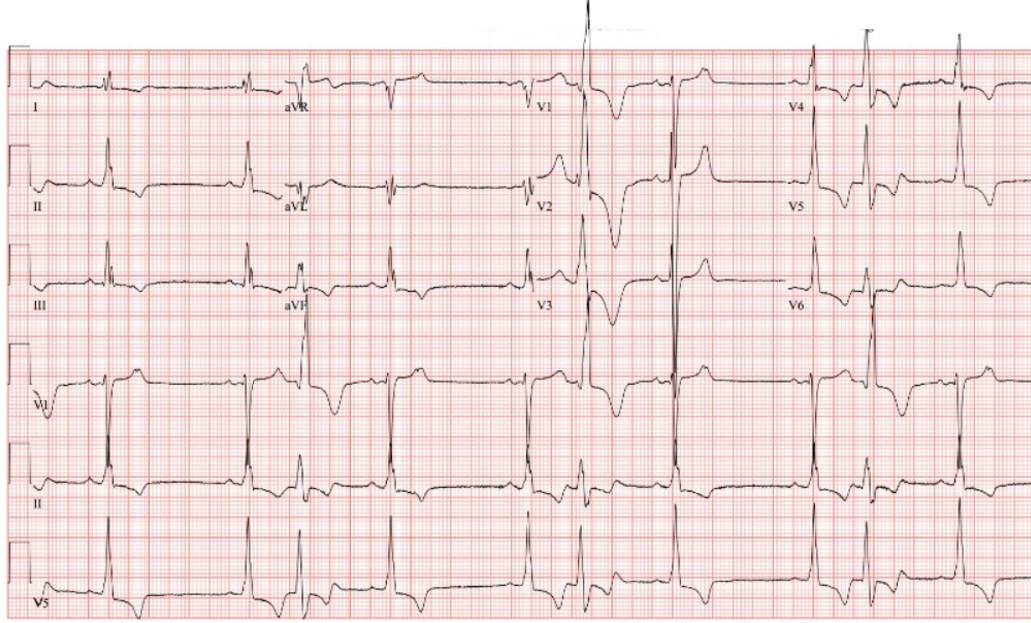
Figure 1
The ECG shows which of the following:
Show Answer
The correct answer is: C. Premature atrial contractions.
The ECG shows sinus rhythm with normal PR interval and prolonged QRS duration (126 ms). The horizontal QRS voltage suggests left ventricular hypertrophy (LVH). The ST-T wave changes are compatible with LVH or a left intraventricular conduction delay. The voltage and ST-T wave changes have been present for over 15 years. The rhythm shows three pauses, all of which are secondary to early premature atrial contractions (PACs) with block at the AV node (red arrows; most readily seen on the T wave of first beat in V1). The third, sixth, and ninth beats are wide and of right bundle branch block. These are proceeded by PACs and are due to ventricular aberration.
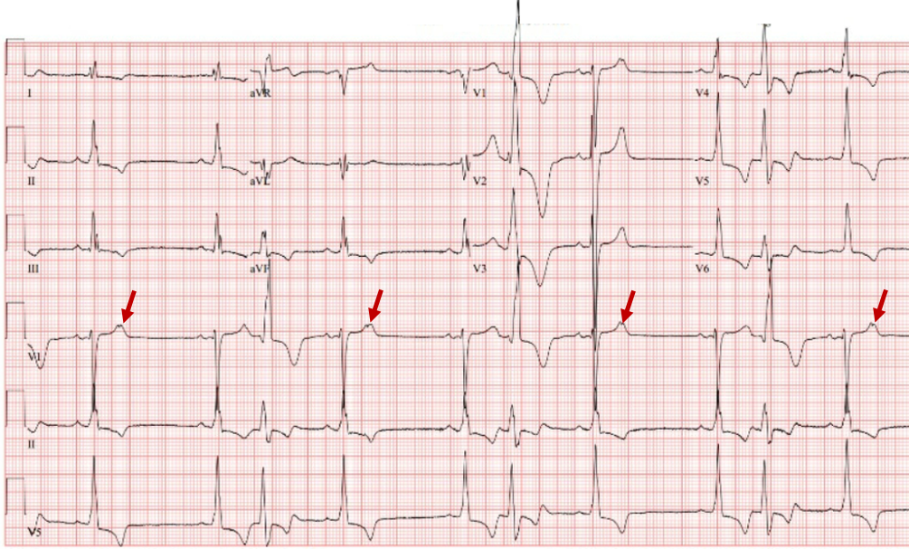
Figure 2
The correct answer is C. No PVCs are present; all rhythm abnormalities are secondary to premature atrial contractions.
References
Spodick DH. Bradycardia due to blocked atrial bigeminy. Am J Geriatr Cardiol 2006;15:328.
Langendorf R. Aberrant ventricular conduction. Am Heart J 1951;41:700-7.