The patient is a 66-year-old male with a history of multiple myeloma, bronchiectasis, chronic dyspnea and end stage renal disease. He reports to the emergency room for cough and two days of respiratory distress.
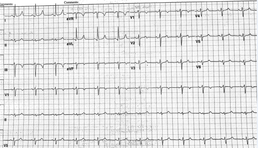
The following ECG is performed:
The correct answer is: D. T wave changes secondary to hyperkalemia.
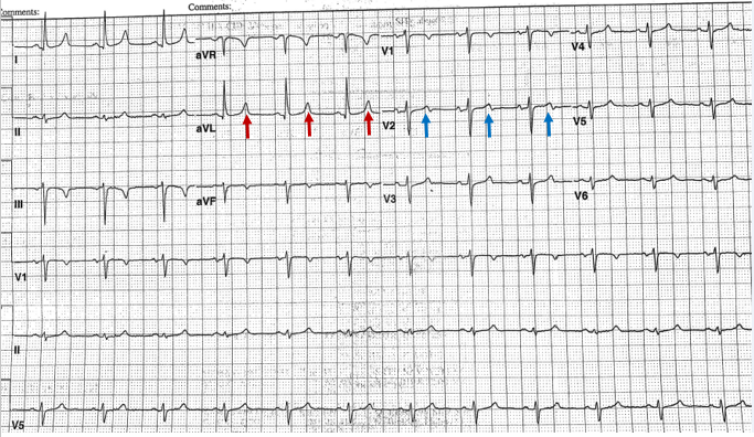
The ECG shows sinus rhythm with a frontal plane axis of approximately -30 degree. The voltage in aVL (12 mm) suggests possible left ventricular hypertrophy, but the voltage in aVL is not reliable in the setting of left anterior fascicular block (LAFB). The poor R wave progression is secondary to LAFB. The patient had an echocardiogram, which showed normal LV function with an EF of 60-65% without hypertrophy. The patient's potassium was 6.1; such is shown in the ECG as a narrow T wave (most noticeable in aVL red arrow) and contributes to the biphasic appearance in V2 (blue arrow).
References
- Wong R, Banker R, Aronowitz P. Electrocardiographic changes of severe hyperkalemia. J Hosp Med 2011;6:240.


