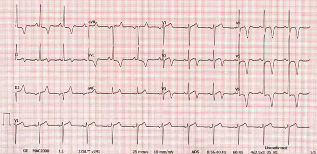
An electrocardiogram is notable for sinus rhythm (Figure 1). Markedly abnormal T waves with diffuse inversions in the limb and precordial leads are present.
Figure 1
Figure 1
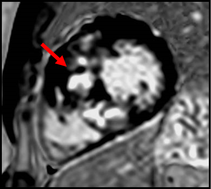
Cardiac magnetic resonance (CMR) imaging reveals increased wall thickness of 24 mm, no obstruction, left ventricular (LV) septal late gadolinium enhancement (LGE)/scar burden in the septum of 18% (red arrow in Figure 2), and normal LV size and systolic function with an ejection fraction (EF) of 68%.
Figure 2
Figure 2
Which one of the following statements is true?
Show Answer
The correct answer is: C. The cardiac magnetic resonance imaging scar burden warrants consideration of implantable cardioverter-defibrillator implantation.
Answer choice A is an incorrect choice. Hypertrophic cardiomyopathy (HCM) and paroxysmal atrial fibrillation are common but, without documentation of atrial fibrillation and its duration, the risks of anticoagulation in this young person outweigh the benefits.
Indications for a primary-prevention implantable cardioverter-defibrillator according to the updated guidelines include:
Family history of sudden death from HCM
Massive LV hypertrophy (>30 mm)
Unexplained syncope
HCM with LV systolic dysfunction (EF <50%)
LV apical aneurysm
Extensive LGE on CMR
Nonsustained ventricular tachycardia (NSVT) on ambulatory monitor
Therefore, answer choice B is an incorrect choice and answer choice C is the correct choice.
Answer D is an incorrect choice because, although one may suspect that NSVT is leading to symptoms, there is not any documentation that this is occurring.
Educational grant support provided by: Bristol Myers Squib To visit the course page for the Hypertrophic Cardiomyopathy: Accelerating Guideline-Driven Care grant, click here.
References
Ommen S, Mital S, Burke M, et al. 2020 ACC/AHA guideline for the diagnosis and treatment of patients with hypertrophic cardiomyopathy: a report of the American College of Cardiology/American Heart Association Joint Committee on Clinical Practice Guidelines. J Am Coll Cardiol 2020;76:3022-55.