A 17-year-old soccer player presents with dyspnea on exertion for two months prior to the visit. There is no personal or family history of syncope or sudden cardiac arrest. In order to evaluate dyspnea an ECG (Figure 1) and echocardiogram (Video Group A & Figure 2) were performed.
Figure 1 (ECG):
Video Group A:
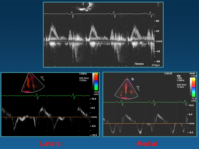
Figure 2:
Based on the above please consider the following question:
What is the most appropriate next step?
Show Answer
The correct answer is: C. Unable to answer, more information required.
The ECG demonstrates sinus rhythm, anterior early repolarization and subtle Q waves which are concerning for pathology including a cardiomyopathy.1-5 Diagnostic criteria for definite left ventricular hypertrophy were absent. The echo shows grossly normal biventricular function, normal valves, and no clear evidence of ventricular hypertrophy. There were abnormal diastolic parameters suggesting an underlying cardiomyopathy.6-7 To further evaluate wall thickness, as well as rule out coronary anomaly and/or scarring, a cardiac MRI was ordered (Video Group B). Massive septal hypertrophy was identified without evidence of myocardial scarring. A diagnosis of hypertrophic cardiomyopathy was made. This case highlights the limitations of echocardiography for evaluation of maximal wall thickness in those with suspected hypertrophic cardiomyopathy. It also underscores the important role cardiac MRI can play in the evaluation of athletes and illustrates the need to consider all modalities when looking for underlying high risk conditions.
Video Group B:
References
Corrado D, Pelliccia A, Heidbuchel H, et al. Recommendations for interpretation of 12-lead electrocardiogram in the athlete. Eur Heart J 2010; 31:243-59.
Uberoi A, Stein R, Perez M, et al. Interpretation of the Electrocardiogram of Young Athletes. Circulation 2011; 124: 746-757.
Drezner JA, Ackerman MJ, Anderson J, et al. Electrocardiographic interpretation in athletes: the 'Seattle Criteria.' Br J Sports Med 2013;47:122-124.
Savage, Seides SF, Clark CE, et al. Electrocardiographic findings in patients with obstructive and nonobstructive hypertrophic cardiomyopathy. Circulation. 1978; 58: 402-408.
Johnson JN, Horner JM, Ackerman MJ, et al. Assessing Electrocardiographic Screening in Elite High School Athletes with Comparison to Adolescents with Hypertrophic Cardiomyopathy. Circulation 2011; 124: A10616.
Pela G, Bruschi G, Montagna L, et al. Left and right ventricular adaptation assessed by Doppler tissue echocardiography in athletes. J Am Soc Echocardiogr 2004;17:205–211.
Kato TS, Noda A, Izawa H, et al. Discrimination of nonobstructive hypertrophic cardiomyopathy from hypertensive left ventricular hypertrophy on the basis of strain rate imaging by tissue Doppler ultrasonography. Circulation 2004;110:3808–3814.