A 45-Year-Old Woman Presenting With Painful Fingers
A 45-year-old woman with hypothyroidism presents to your office complaining of painful fingers. When exposed to cold temperatures, the fingers on her right hand become markedly cold, pale, stiff, and achy. Upon questioning, she denies involvement of her thumbs, fingers on her left hand, or toes, and the skin changes do not extend beyond the digits. She has noticed these symptoms for the past two years, but they have increased in severity this winter, as she has been forced to spend more time shoveling snow. Her symptoms resolve with warming, and she has been wearing gloves, even while indoors, to help prevent attacks. There is no skin breakdown or discoloration that persists after warming. She denies any tobacco or illicit drug use or family members with similar symptoms.
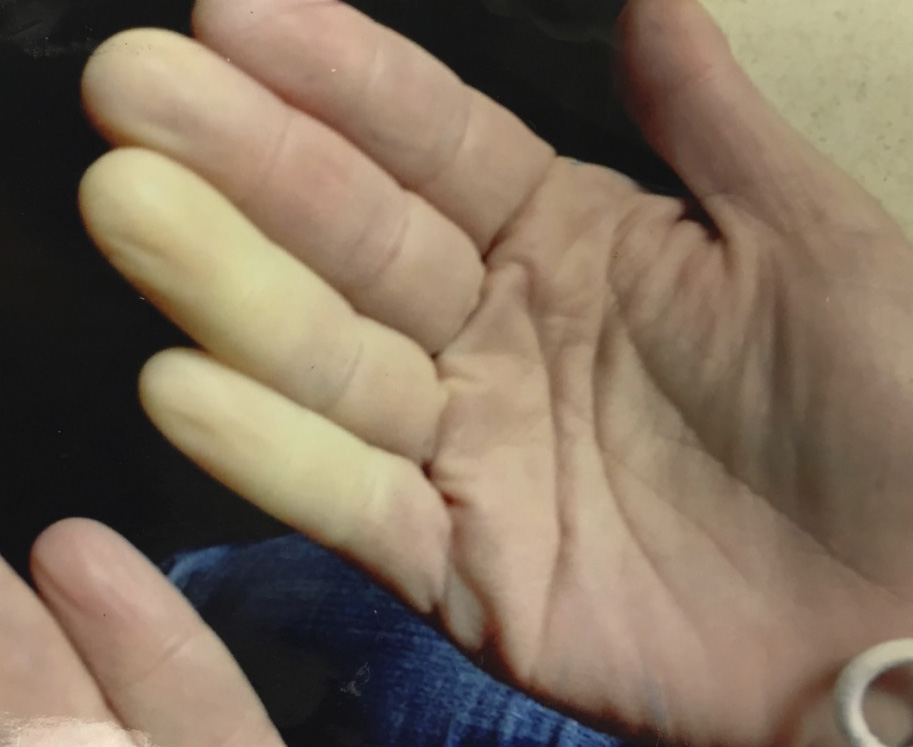
On exam, there is pallor of the third, fourth, and fifth digits of the right hand (Figure 1), and these digits are cold to touch with sluggish capillary refill. The digits of the left hand are unaffected. After she wraps her hands in a warm blanket for several minutes, the digits return to normal. Nailfolds are smooth and uniformly pink. The skin is intact without sclerodactyly. Radial and ulnar arterial pulses are 2+ bilaterally.
Figure 1
Given the description and physical exam, which of the following describes the most likely diagnosis?
Show Answer
The correct answer is: D. Secondary Raynaud's phenomenon.
Raynaud's phenomenon is defined by the presence of well-demarcated, cold-induced digital pallor, cyanosis, and/or pain that completely resolves with warming. It affects the fingers, toes, or occasionally nose, tip of the tongue, or earlobe.1 There are three phases of a Raynaud's "attack." During the initial phase, vasospasm of small arteries and arterioles causes hypoperfusion and pallor of the affected area after cold exposure or an emotional stressor.2 As the residual blood begins to desaturate, the skin becomes blue in the cyanotic phase.1 Eventually, during the final phase, resolution of vasoconstriction causes hyperemia of the digits. Patients experience pain or paresthesias due to soft tissue, muscle, and small nerve fiber ischemia. Estimates of the prevalence of Raynaud's phenomenon vary widely and can be confused with reports of general cold sensitivity. However, survey analyses yield a prevalence of up to 14% in men and 20% in women.3 Interestingly, the prevalence varies widely in different geographic regions with a decreased prevalence in warm climates.4,5
When evaluating Raynaud's phenomenon, it is important to distinguish between primary and secondary cases. Primary Raynaud's phenomenon typically develops before age 30.6 It presents as symmetric involvement of the digits without tissue necrosis or ulceration.7 Nailfold capillaries appear normal, and there are no signs or symptoms suggestive of a secondary cause.2 The erythrocyte sedimentation rate as well as antinuclear antibody testing are also normal. Because of this patient's age and the asymmetric nature of her symptoms, answer option C is incorrect.
In contrast, patients usually develop secondary Raynaud's phenomenon over the age of 40 years, as in the patient from this Patient Case Quiz.6 Unlike primary Raynaud's phenomenon, patients can develop asymmetric digital involvement, prolonged symptoms, and ulceration.2 Nailfold capillaroscopy often reveals abnormal or absent capillary loops, which is indicative of an underlying connective tissue disease.8 If this is not available, bedside examination of the nailfolds is also useful. Rather than a contiguous strip of pinkish tissue, nailfolds in patients with Raynaud's phenomenon may exhibit intermixed white areas consistent with damaged or absent capillaries. Some common causes of secondary Raynaud's phenomenon are listed in Table 1. Findings from the history and physical examination that suggest a secondary cause should prompt disease-specific diagnostic testing.
Table 1: Causes of Secondary Raynaud's Phenomenon
|
Connective tissue disorders – systemic sclerosis, CREST syndrome, systemic lupus erythematosus |
|
Malignancy |
|
Hypothyroidism |
|
Vibration or thermal injury – heavy machinery use, frostbite |
|
Malignancy |
|
Drugs or toxins – chemotherapeutic agents, ergots |
|
Arterial occlusive disease – atherosclerosis, thromboangiitis obliterans |
|
Hematologic disorders – cryoglobulinemia, cold agglutinin disease |
The pathophysiology of Raynaud's phenomenon remains incompletely understood and can vary widely among individuals. In primary Raynaud's phenomenon, it seems to be caused by an exaggeration of the normal vasoconstrictive response.2 However, particularly in secondary cases, there may be some degree of endothelial activation and damage in addition to perturbations of α-adrenergic signaling.9 Symptoms often cluster within families, suggesting genetic factors may also play a role.10
Cold avoidance, protection of exposed skin, and smoking cessation are important components of therapy in all patients with Raynaud's phenomenon.9 In individuals with ongoing pain or tissue necrosis, pharmacologic therapy should be initiated in addition to treatment of the underlying disorder in secondary cases. Nifedipine, a dihydropyridine calcium channel blocker, is typically used as first-line therapy and has proven beneficial in placebo-controlled trials.11 Topical nitrates can be effective in patients who experience symptoms despite calcium channel blocker therapy, although systemic absorption may cause symptoms including headache, flushing, nausea, and hypotension.2 The α1-antagonist prazosin is also beneficial in many patients.12 Other potential pharmacologic options include losartan, fluoxetine, sildenafil, and intravenous prostaglandins.2,13 There are some data to support digital artery sympathectomy, but this should be reserved for cases refractory to standard medical therapy.14
Because clinicians frequently encounter a variety of conditions involving pain or discoloration of the digits, one must be able to distinguish Raynaud's phenomenon from other clinical entities. Erythromelalgia is characterized by severe pain, erythema, and localized edema of the digits or extremities. It is thought to be a neurovascular disorder caused by an imbalance in constriction and dilation of arterioles and venules, potentially mediated by a small fiber neuropathy.15 In contrast to Raynaud's phenomenon, the pain is exacerbated by warmth and relieved by exposure of the affected area to cold, making answer option A incorrect. Many patients use ice water baths or fans for relief. Recently, some data suggest a role for topical amitriptyline and ketamine in managing the pain associated with erythromelalgia.16
Acrocyanosis is a syndrome notable for painless, bluish discoloration of the hands, feet, or portions of the face.17 Cold exposure often triggers this discoloration, but it typically persists despite rewarming. Patients may also note hyperhidrosis of the affected area. Although acrocyanosis usually does not require therapy, it can co-exist with Raynaud's phenomenon. However, because our patient notes only pallor and painful digits that fully resolve with rewarming, answer option B is incorrect.
Thromboangiitis obliterans, or Buerger's disease, is a small- and medium-vessel vasculitis that affects the arteries, veins, and nerves of the extremities.18 Patients often present with claudication and frank tissue loss due to progressive occlusion of small vessels. Tobacco exposure, including smokeless tobacco, is the key component in the development of this disease, and cessation of tobacco use is the primary therapy. Of note, up to 40% of patients with thromboangiitis obliterans may also exhibit features of Raynaud's phenomenon.19 Given the intermittent nature of the patient's symptoms in addition to the lack of tobacco exposure, answer option E is incorrect.
References
- Cooke JP, Marshall JM. Mechanisms of Raynaud's disease. Vasc Med 2005;10:293-307.
- Wigley FM. Clinical practice. Raynaud's Phenomenon. N Engl J Med 2002;347:1001-8.
- Silman A, Holligan S, Brennan P, et al. Prevalence of symptoms of Raynaud's phenomenon in general practice. BMJ 1990;301:590-2.
- Maricq HR, Weinrich MC, Keil JE, et al. Prevalence of Raynaud phenomenon in the general population. A preliminary study by questionnaire. J Chronic Dis 1986;39:423-7.
- Maricq HR, Carpentier PH, Weinrich MC, et al. Geographic variation in the prevalence of Raynaud's phenomenon: a 5 region comparison. J Rheumatol 1997;24:879-89.
- Planchon B, Pistorius MA, Beurrier P, et al. Primary Raynaud's phenomenon: age of onset and pathogenesis in a prospective study of 424 patients. Angiology 1994;45:677-86.
- LeRoy EC, Medsger TA Jr. Raynaud's phenomenon: a proposal for classification. Clin Exp Rheumatol 1992;10:485-8.
- Kallenberg CG. Early detection of connective tissue disease in patients with Raynaud's phenomenon. Rheum Dis Clin North Am 1990;16:11-30.
- Block JA, Sequeira W. Raynaud's phenomenon. Lancet 2001;357:2042-8.
- Freedman RR, Mayes MD. Familial aggregation of primary Raynaud's disease. Arthritis Rheum 1996;39:1189-91.
- Belch JJF, Ho M. Pharmacotherapy of Raynaud's phenomenon. Drugs 1996;52:682-95.
- Pope J, Fenlon D, Thompson A, et al. Prazosin for Raynaud's phenomenon in progressive systemic sclerosis. Cochrane Database Syst Rev 2000;2:CD000956.
- Fries R, Shariat K, von Wilmowsky H, Bohm M. Sildenafil in the treatment of Raynaud's phenomenon resistant to vasodilator therapy. Circulation 2005;112:2980-5.
- Flatt AE. Digital artery sympathectomy. J Hand Surg 1980;5:550-6.
- Cohen JS. Erythromelalgia: New theories and new therapies. J Am Acad Dermatol 2000;43:841-7.
- Poterucha TJ, Weiss WT, Warndahl, RA, et al. Topical amitriptyline combined with ketamine for the treatment of erythromelalgia: a retrospective study of 36 patients at Mayo Clinic. J Drugs Dermatol 2013;12:308-10.
- Kurklinsky AK, Miller VM, Rooke TW. Acrocyanosis: the Flying Dutchman. Vasc Med 2011;16:288-301.
- Piazza G, Creager MA. Thromboangiitis Obliterans. Circulation 2010;121:1858-61.
- Olin JW. Thromboangiitis obliterans (Buerger's disease). N Engl J Med 2000;343:864-9.