An asymptomatic 3-year-old boy presents to the pediatrician's office for a routine well child visit. On physical exam, he has normal vitals with no dysmorphic features. The precordium is quiet with normal heart sounds. A grade I out of VI soft systolic ejection murmur is heard at the base. A grade III out of IV high-frequency holodiastolic murmur is heard at the left midsternal border radiating to the apex. Pulses are prominent but equal. There is no sternal deformity, hypermobility, skin striae, or abnormal scarring. There is no family history of congenital heart disease, cardiomyopathy, connective tissue disorder, sudden cardiac arrest, or sudden death. His chest X-ray is shown in Figure 1. Select images from the echocardiogram (echo) are shown in Videos 1, 2, 3 and 4. Computed tomography (CT) angiography three-dimensional reconstruction is shown in Video 5.
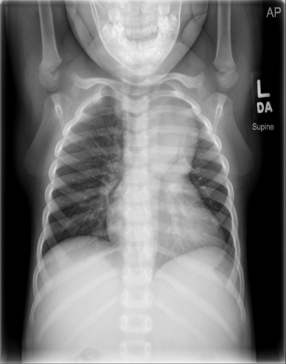
Figure 1: Chest X-Ray
Video 1: Echo: Parasternal Long-Axis View
Video 2: Echo: Suprasternal View
Video 3: Parasternal Short-Axis View
Video 4: Echo: Parasternal Long-Axis View
Video 5: CT Angiogram of Entire Aorta
Which of the following tests are recommended for the child and other family members to identify the etiology of his condition and its long-term prognosis?
Show Answer
The correct answer is: C. Echos for all first-degree family members; ophthalmological evaluation and genetic testing for connective tissue panel in the child.
The chest X-ray in Figure 1 shows an enlarged mediastinal silhouette suspicious for ascending aorta pathology. The echo images in Videos 1 and 2 show severe dilation of the ascending aorta, consistent with a thoracic aortic aneurysm (maximum diameter 43 mm = Z score of 11.5) with no evidence of aortic dissection. The proximal ascending aorta is moderately dilated with a maximum diameter of 25 mm at the sinus of valsalva. Videos 3 and 4 show a trileaflet aortic valve with mild-to-moderate insufficiency. CT angiography with three-dimensional reconstruction in Video 5 confirms the echo findings and reveals normal descending aorta dimensions.
Thoracic aortic aneurysms in childhood are phenotypically and genotypically heterogeneous, and may be classified into non-syndromic and syndromic types. Familial thoracic aorta aneurysm and dissection (FTAAD) is not an infrequent cause of aortic aneurysms in young persons without overt syndromic features. The majority of the mutations causing FTAAD are autosomal dominant with complete or incomplete penetrance, depending on the underlying genetic mutation. There is variability in the age of onset of aortic dilation; thus, lifelong monitoring of patients with a positive gene mutation is recommended. Syndromic aortic aneurysms may be due to Marfan (FBN1 mutations), Loeys-Dietz (TGFBRI and TGFBRII mutations) and multisystemic smooth muscle dysfunction (MSMD; ACTA2 R159H mutations) syndromes. In addition to aortic diameter, the underlying gene defect plays an important role in the frequency of aortic dissection, enabling important clinical management risk stratification. In this case, it is important to determine the underlying etiology of aortic dilation to assess the risk of dissection and subsequent aneurysm formation, and to provide counseling regarding other at-risk family members. Ophthamologic evaluation is important to assess for ocular features of Marfan (myopia, lens subluxation) and MSMD syndrome (iris flocculi, congenital mydriasis). With the aortic diameter remaining the primary risk predictor of aortic dissection, it is important for the family members to obtain screening echos.
References
Elisabeth Gillis, Lut Van Laer, Bart L. Loeys. Genetics of thoracic aortic aneurysm: at the crossroad of transforming growth factor-β signaling and vascular smooth muscle cell contractility. Circ Res 2013;113:327-340.
Landis BJ, Ware SM, James J, Shikany MS, Martin LJ, Hinton RB. Clinical stratification of pediatric patients with idiopathic thoracic aorta aneurysm. J Pediat 2015:167:131-7.
Pierpont ME, Lacro RV. Children with thoracic aortic aneurysm: challenges in diagnosis and therapy. J Pediatr 2015 167:14-6.