A Case of a Healthy, Athletic Teenager Who Presents With Isolated Symptoms of Exercise-Induced Dyspnea | Patient Case Quiz
A 16-year-old athletic female who has a past medical history of asthma requiring bronchodilators as needed, presents with shortness of breath with exercise. Symptoms usually occur 15 minutes after exercise, are worse with intense physical activity, and cause her to stop running.
There is no history of associated wheezing or stridor. There were no environmental triggers or history of recent infections. There was also no history of palpitations or syncope.
She was diagnosed with exercise-induced asthma two years ago and was prescribed an albuterol inhaler without significant benefit. On examination, she is a relatively thinly built female with a body mass index (BMI) of 22 kg/m2. Chest and cardiac auscultation are normal. Electrocardiogram (ECG) reveals first-degree heart block and echocardiogram demonstrates normal biventricular size and function.
Spirometry and lung volume reveal the following data (Table 1).
Table 1
|
|
Actual |
% Predicted |
|
FVC |
3.07L |
92 |
|
FEV1 |
2.89L |
97 |
|
FEV1/FVC |
94 |
|
|
FEF 25/75 |
4.3L |
115 |
|
FRC |
2.22L |
94 |
|
RV |
1.08L |
84 |
|
TLC |
4.19L |
90 |
|
RV/TLC |
26 |
93 |
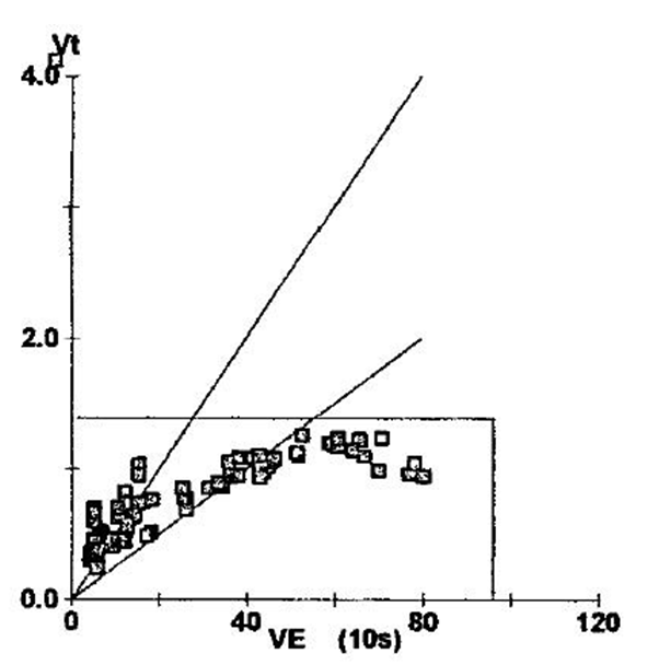
She undergoes a cardiopulmonary exercise stress test with incremental increase in either speed or incline every minute as tolerated. During the test, breath-by-breath analysis of oxygen consumption (VO2), CO2 production (VCO2), tidal volume (VT) and minute ventilation (VE) are obtained. Also, pre- and post-exercise spirometry are done and showed no significant decreased in FEV1 after exercise. Peak cardiovascular and ventilatory responses are outlined in Table 2.
Table 2
|
|
Predicted |
Measured |
% Predicted |
|
VO2 (L/min) |
1.94 |
2.38 |
123 |
|
VCO2 (L/min) |
1.86 |
2.27 |
122 |
|
Anaerobic Threshold (AT) |
>0.777 |
1.94 |
|
|
Heart Rate (bpm) |
188 |
164 |
87 |
|
O2 Pulse |
10.6 |
14.5 |
137 |
|
VE Max (L/min) |
96.8 |
81.5 |
84 |
|
VT (L) |
1.38 |
0.99 |
72 |
|
VT/FVC |
0.6 |
0.33 |
54% |
|
Respiratory Rate (RR) |
<50 |
79 |
|
|
Breathing Reserve (%) |
20-40 |
15 |
|
On a follow-up evaluation, pulmonary function tests continue to be normal with persistent symptoms. She undergoes maximal inspiratory pressure (MIP) and maximal expiratory pressure (MEP) measurements that reveal the following results (Table 3).
Table 3
|
|
Predicted |
Measured |
% Predicted |
|
PImax/MIP(cmH2O) |
-75.4 |
-46.13 |
61 |
|
PEmax/MEP(cmH2O) |
100.8 |
45.34 |
45 |
Which of the following statements best explains the cause of this patient's symptoms?
Show Answer