A 55 year old man with paroxysmal atrial fibrillation (AF) was referred for catheter ablation because of symptomatic recurrent episodes (2-3 episodes per month lasting between 2-6 hours) despite multiple antiarrhythmic drugs trials (including amiodarone).
No hypertension, diabetes or coronary heart disease.
Normal echocardiogram.
Magnetic resonance imaging (MRI) demonstrated normal pulmonary vein anatomy with 4 distinct pulmonary vein ostia.
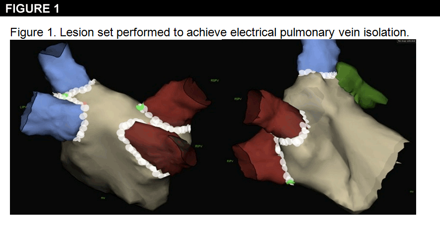
Pulmonary vein isolation (PVI) was performed using an irrigated-tip radiofrequency ablation catheter guided by a circular mapping catheter. A three-dimensional, nonfluoroscopic mapping system (NavX system) was used for the procedure (Figure 1).
Spontaneous initiation of AF from the left superior pulmonary vein (LSPV) was observed during the case.
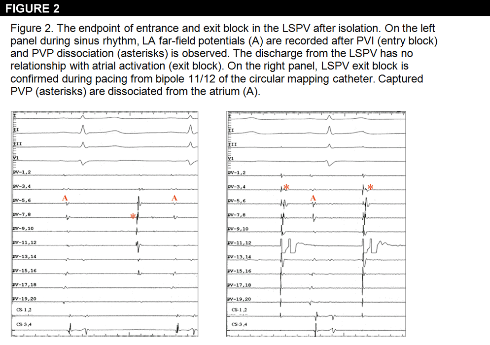
The endpoint of electrical pulmonary vein isolation (entrance and exit block) was confirmed in each of the 4 PVs after an observation time of 30 minutes post ablation (Figure 2).
At that point in the ablation procedure, the next step that may help to ensure durable PV isolation and avoid PV reconnection would be:
Show Answer
The correct answer is: C. Adenosine IV bolus injections to look for dormant PV conduction
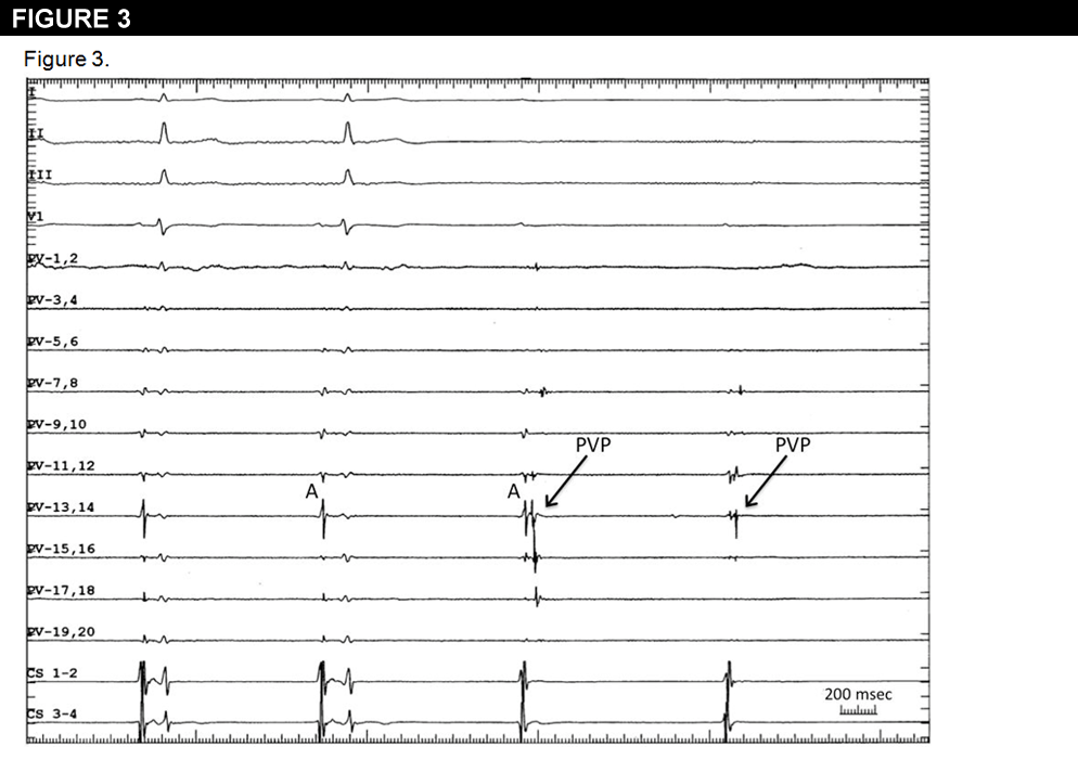
After isolation of the LSPV, no pulmonary vein potentials (PVP) are observed during the first two beats, with the circular mapping catheter (PV 1,2 to PV 19,20) recording only atrial far-field signals (A). Adenosine administration (12 mg IV bolus) results in AV block with transient recovery of PV conduction. The atrial far-field signal (A) is recorded first, followed by PVP (Figure 3).
PVI is an effective treatment for AF. Nevertheless, many patients require repeated ablation procedures because of AF-recurrence, in most cases associated with reconnection of previously-isolated PVs.
The appearance of PV-atrial conduction block during the initial procedure may be permanent or indicate dormant conduction, which subsequently recovers. Injecting IV adenosine to restore conduction in viable but acutely non-conducting PVs may distinguish permanent block from dormant PV conduction, thereby allowing additional targeted ablation.(1) Adenosine activates outward K+-currents and leads to cellular hyperpolarization, especially in atrial cells. This hyperpolarization increases INa by removing voltage-dependent inactivation, revealing dormant conduction in viable PVs.(2)
Adenosine is superior to isoproterenol in revealing dormant PV clinically and experimentally, due to greater adenosine-induced hyperpolarization.(3)
A longer observation period after PVI has been proposed to assess spontaneous recovery of PV-atrial conduction. However, this approach is impractical and adenosine has been shown to reveal additional PV reconnections.(4-5)
A few studies have reported the potential benefit of using intravenous adenosine to unmask dormant PV conduction.(4-7) In these studies, dormant conduction was present in 41-60% of patients after PVI. Comparing adenosine-guided PVI to historical control groups without adenosine-guided ablation, a reduction in AF recurrence was noted. Although these retrospective studies are promising, a prospective randomized controlled trial is currently being performed to definitely address the critical question of whether ablation at sites of adenosine-induced transient PV re-conduction improves outcomes after PVI.(8)
References
Arentz T, Macle L, Kalusche D, et al. "Dormant" pulmonary vein conduction revealed by adenosine after ostial radiofrequency catheter ablation. J Cardiovasc Electrophysiol 2004;15:1041-7.
Datino T, Macle L, Qi X, et al. Mechanisms by which adenosine restores conduction in dormant canine pulmonary veins. Circulation 2010;121:963-72.
Datino T, Macle L, Chartier D et al. Differential effectiveness of pharmacological strategies to reveal dormant pulmonary vein conduction: a clinical-experimental correlation. Heart Rhythm 2011;8:1426-33.
Yamane T, Matsuo S, Date T et al. Repeated provocation of time- and ATP-induced early pulmonary vein reconnections after pulmonary vein isolation: eliminating paroxysmal atrial fibrillation in a single procedure. Circ Arrhythm Electrophysiol 2011;4:601-8.
Ninomiya Y, Iriki Y, Ishida S, et al. Usefulness of the adenosine triphosphate with a sufficient observation period for detecting reconduction after pulmonary vein isolation. Pacing Clin Electrophysiol 2009;32:1307-12.
Matsuo S, Yamane T, Date T, et al. Reduction of AF recurrence after pulmonary vein isolation by eliminating ATP-induced transient venous re-conduction. J Cardiovasc Electrophysiol 2007;18:704-8.
Hachiya H, Hirao K, Takahashi A et al. Clinical implications of reconnection between the left atrium and isolated pulmonary veins provoked by adenosine triphosphate after extensive encircling pulmonary vein isolation. J Cardiovasc Electrophysiol. 2007;18:392-398.
Macle L, Khairy P, Verma A et al. Adenosine Following Pulmonary Vein Isolation to Target Dormant Conduction Elimination (ADVICE): Methods and Rationale. Can J Cardiol 2012;[Epub ahead of print].