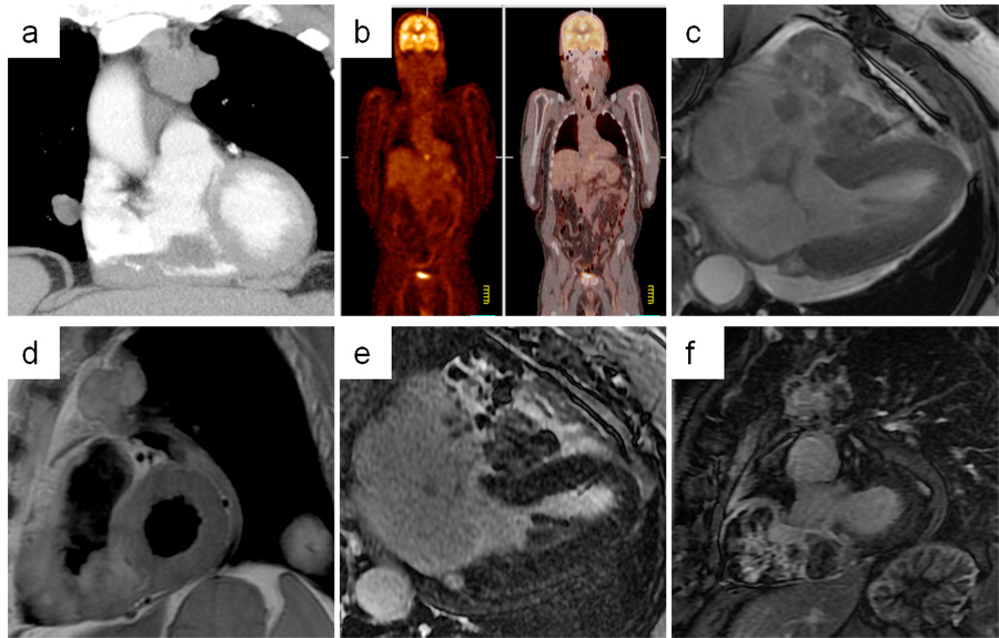
A 73-year-old man with a history of metastatic malignant melanoma was referred for the assessment of a cardiac mass that was detected on surveillance computed tomography scan (Figure a).
|
Figure (a-f)
|
|
|
Review of a previous positron emission tomography scan showed a hypermetabolic focus in the area (
Figure b). Transesophageal echocardiography revealed a large echodense right ventricular mass (
Video 1). Cardiac magnetic resonance (CMR) imaging was performed. CMR steady state free precession (SSFP) cine images (
Videos 2 and 3; Figure c), as well as T1-weighted (
Figure d) and late gadolinium images (
Figures e-f) are shown.
The correct answer is: D. Hyperintensity of the mass on T1-weighted CMR sequences is consistent with the diagnosis of a cardiac melanoma metastasis.
The mass shows features characteristic of a metastatic deposit of melanoma in the RV. Infiltration of the RV free wall indicates an invasive process; patchy late gadolinium enhancement confirms a degree of vascularity allowing gadolinium to perfuse the mass, and also points to necrosis/fibrosis within the mass. The hyperintensity on T1-weighted images both in the mass and the pulmonary metastases (Figure d) is suggestive of melanin content - as melanin is known to shorten the T1 relaxation time due to paramagnetic metal scavenging.(1) Inflammation and oedema is identified using T2 weighted sequences.
Ventricular thrombi do not have gadolinium uptake, and do not demonstrate late gadolinium enhancement (unless in rare organised and vascularised chronic thrombi), and their signal characteristics on T1-weighted images depends on the age – acute and subacute ones are bright, whereas chronic organized thrombi have low signal intensity due to depleted water with or without calcification of the thrombus.(2)
Histopathology was confirmatory of metastatic melanoma in this case.
References
- 1. Enochs WS, Petherick P, Bogdanova A, Mohr U, Weissleder R. Paramagnetic metal scavenging by melanin: MR imaging. Radiology 1997; 204:417- 23
- 2. Patrick J. Sparrow, John B. Kurian, Tim R. Jones, Mohan U.Sivananthan. MR Imaging of Cardiac Tumors. RadioGraphics 2005; 25:1255-1276.