A 63-year-old female patient presented to the outpatient cardiovascular clinic for evaluation of a 4-month history of exertional chest pain. She described the chest pain as a moderate-intensity, intermittent chest discomfort that occurs with exertion and resolves with rest or use of sublingual nitroglycerin. Her medical history was significant for hypertension and dyslipidemia. On examination, her blood pressure was 132/68 mmHg, and her heart rate was 65 bpm. Her lipid profile showed low-density lipoprotein of 84 mg/dL, high-density lipoprotein of 43 mg/dL, and triglyceride of 103 mg/dL. Her medication regimen included 81 mg of aspirin daily, nitroglycerin, and 20 mg of atorvastatin daily. Based on her symptoms, she underwent a coronary angiogram several weeks ago, which showed mild-to-moderate diffuse, nonobstructive coronary artery disease (CAD).
Due to the concern for microvascular dysfunction, an adenosine stress cardiac magnetic resonance imaging (CMRI) test was ordered. The CMRI showed that her left ventricular size and systolic function were normal with a left ventricular ejection fraction (LVEF) of 68%. There was mild tricuspid regurgitation, moderate aortic stenosis with peak aortic valve velocity of 2.4 ms, and mild aortic regurgitation. First-pass stress perfusion imaging showed diffuse, circumferentially abnormal subendocardial perfusion prominent at the base and consistent with microvascular dysfunction. No wall motion abnormalities were detected. No significant scar/fibrosis was detected on late postgadolinium enhancement imaging (Figure 1 and Videos 1-6). Her CMRI appeared to be consistent with microvascular dysfunction.
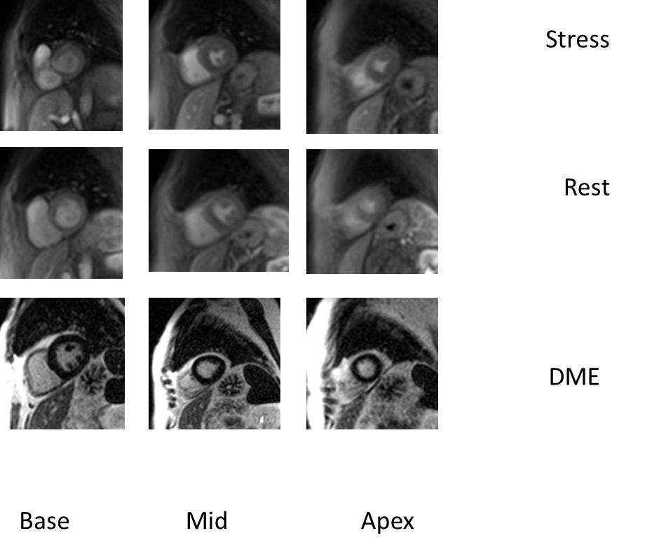
Figure 1
This image shows stress and rest adenosine stress perfusion imaging with diffuse subendocardial perfusion abnormality in the basal and mid segments of the left ventricle myocardium (140 mcg/kg of adenosine over 4 min with multislice stress perfusion using 0.075 mmol/kg intravenous gadolinium diethylenetriaminepentaacetic acid). The rest imaging was obtained 15 min post stress imaging. The delayed myocardial enhancement imaging was conducted 5-10 min after final contrast administration.
Video 1: Rest perfusion image of apical left anterior descending (LAD).
Video 2: Rest perfusion image of mid-LAD with diffuse subendocardial perfusion abnormality.
Video 3: Rest perfusion image of basal LAD with diffuse subendocardial perfusion abnormality.
Video 4: Stress perfusion image of apical LAD.
Video 5: Stress perfusion image of mid-LAD with diffuse subendocardial perfusion abnormality.
Video 6: Stress perfusion image of basal LAD with diffuse subendocardial perfusion abnormality.
What is your opinion regarding her long-term CAD prognosis given the abnormal CMRI perfusion results?
Show Answer
The correct answer is: B. The presence of a subendocardial perfusion defect confers a worse long-term prognosis.
Case Explanation:
Stress CMRI has a role in both the diagnosis and prognostication of CAD. Pharmacologic stress is the frequently used stress modality due to incompatibility of standard exercise equipment with MRI and the difficulty associated with performing CMRI with postexercise tachypnea and tachycardia. However, a modified treadmill stress CMRI protocol has been developed and validated.1 Stress CMRI has the advantage of being able to evaluate several prognostic parameters within a single imaging test: wall motion abnormality, perfusion defect, myocardial scar/fibrosis, and ejection fraction.
A meta-analysis of 14 studies (12,178 patients) with a weighted mean follow-up of 25.3 months showed that a normal stress CMRI had a 98.1% negative predictive value (NPV) (95% confidence interval [CI], 97.26-98.83) for nonfatal myocardial infarction (MI) and cardiac death.2 In this meta-analysis, the absence of inducible perfusion defect and the absence of inducible wall motion abnormality had similar NPV for major coronary events (98.39% vs. 97.31%, respectively; p = .227 by meta-regression analysis). Another meta-analysis involving 19 studies (11,636 patients with known or suspected CAD) with a mean follow-up of 32 months showed that patients with a positive stress CMRI had a higher combined outcome (cardiovascular death and nonfatal MI) annual event rate of 4.9% versus 0.8% for a negative stress CMRI (p < 0.0001), 2.8% versus 0.3% for cardiovascular death (p < 0.0001), and 2.6% versus 0.4% for MI (p < 0.0005).3
Regional wall motion abnormalities and perfusion defects detected during stress CMRI have been shown to have a complementary prognostic effect and enhance CMRI sensitivity for ischemia detection.4,5 The detection of myocardial scar/fibrosis by late gadolinium enhancement (LGE) has also been shown to have complementary prognostic value in combination with perfusion and wall motion evaluation during stress CMRI.6,7 The incremental prognostic value of abnormal perfusion and abnormal wall motion was demonstrated in a recent study8 that evaluated the prognostic value of stress CMRI in patients with known or suspected CAD. Seven hundred and ninety-three consecutive patients with chest pain were studied. Patients were classified in three groups: group 1 (no reversible ischemia), group 2 (stress perfusion defect alone), and group 3 (stress perfusion defect plus abnormal wall motion). The end points were "all cardiac events" (MI, cardiac death, and revascularization) and "hard cardiac events" (all cardiac events excluding revascularization). During the 810 ± 665 day follow-up, the hard event rate was 4% in patients with a negative stress CMRI (group 1); 8% in patients with a positive stress CMRI for perfusion defect alone (group 2), and 21% in patients with perfusion defect plus abnormal wall motion (group 3). In univariate analysis, the hazard ratio (HR) for hard cardiac events was 1.99 (95% CI, 1.45-2.74; p < 0.001) in group 2 and 2.58 (95% CI, 1.50-4.45; p < 0.001) in group 3. Multivariate analysis revealed that the LGE and stress perfusion defect plus abnormal wall motion were independent predictors of all and hard cardiac events. Another pharmacologic stress CMRI study involving 149 patients with known or suspected CAD9 showed that the presence of different CMRI parameters significantly predicted major adverse cardiovascular events: presence of perfusion defects [HR 6.4; 95% CI, 2.25-18.2; p = 0.001]; LVEF < 40% (HR 6.4; 95% CI, 2.28-18.5; p = 0.001), LGE (HR 3.45; 95% CI, 1.33-8.95; p = 0.011), and any resting regional wall motion abnormality (HR 3.12; 95% CI, 1.10-8.88, p = 0.033). The absence of perfusion defects revealed an NPV of 96%. However, the absence of perfusion defects in addition to the absence of other resting CMRI abnormalities (LVEF < 40%, regional wall motion abnormalities, and LGE) improved the NPV from 96 to 99%.
One stress CMRI study demonstrated that the lowest annual event rate (cardiac death or nonfatal MI) was seen in patients without reversible perfusion defects or LGE.10 Another study showed that a very low annual cardiac mortality was seen in patients without perfusion defect, LGE, normal LVEF, and normal aortic flow.11
Answers A, C, D, and E are incorrect. Wall motion abnormality, LVEF, perfusion defect, and myocardial scar/fibrosis can be used as prognostic parameters in CAD. In the ischemic cascade, perfusion abnormality precedes diastolic and systolic dysfunction and is a more sensitive indicator of myocardial ischemia. This patient was initiated on a calcium channel blocker, and her statin dose was increased. Her chest pain improved significantly with this regimen. It is important to remember that patient's pretest cardiac risk assessment affects the prognostic ability of stress CMRI.
References
Raman SV, Dickerson JA, Jekic M, et al. Real-time cine and myocardial perfusion with treadmill exercise stress cardiovascular magnetic resonance in patients referred for stress SPECT. J Cardiovasc Magn Reson 2010;12:41.
Gargiulo P, Dellegrottaglie S, Bruzzese D, et al. The prognostic value of normal stress cardiac magnetic resonance in patients with known or suspected coronary artery disease: a meta-analysis. Circ Cardiovasc Imaging 2013;6:574-82.
Lipinski MJ, McVey CM, Berger JS, Kramer CM, Salerno M. Prognostic value of stress cardiac magnetic resonance imaging in patients with known or suspected coronary artery disease: a systematic review and meta-analysis. J Am Coll Cardiol 2013;62:826-38.
Gebker R, Jahnke C, Manka R, et al. Additional value of myocardial perfusion imaging during dobutamine stress magnetic resonance for the assessment of coronary artery disease. Circ Cardiovasc Imaging 2008;1:122-30.
Korosoglou G, Elhmidi Y, Steen H, et al. Prognostic value of high-dose dobutamine stress magnetic resonance imaging in 1,493 consecutive patients: assessment of myocardial wall motion and perfusion. J Am Coll Cardiol 2010;56:1225-34.
Flett AS, Westwood MA, Davies LC, Mathur A, Moon JC. The prognostic implications of cardiovascular magnetic resonance. Circ Cardiovasc Imaging 2009;2:243-50.
Zemrak F, Petersen SE. Late gadolinium enhancement CMR predicts adverse cardiovascular outcomes and mortality in patients with coronary artery disease: systematic review and meta-analysis. Prog Cardiovasc Dis 2011;54:215-29.
Pontone G, Andreini D, Bertella E, et al. Prognostic value of dipyridamole stress cardiac magnetic resonance in patients with known or suspected coronary artery disease: a mid-term follow-up study. Eur Radiol 2015 Oct 29 [Epub ahead of print].
Freed BH, Narang A, Bhave NM, et al. Prognostic value of normal regadenoson stress perfusion cardiovascular magnetic resonance. J Cardiovasc Magn Reson 2013;15:108.
Steel K, Broderick R, Gandla V, et al. Complementary prognostic values of stress myocardial perfusion and late gadolinium enhancement imaging by cardiac magnetic resonance in patients with known or suspected coronary artery disease. Circulation 2009;120:1390-400.
Bingham SE, Hachamovitch R. Incremental prognostic significance of combined cardiac magnetic resonance imaging, adenosine stress perfusion, delayed enhancement, and left ventricular function over preimaging information for the prediction of adverse events. Circulation 2011;123:1509-18.