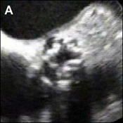
A 79-year-old woman with known severe aortic stenosis and concomitant moderate aortic regurgitation underwent electrocardiogram-gated cardiac computed tomography angiography in addition to TEE for planning of trancatheter aortic valve implantation, which was preferred to conventional valve replacement with sternotomy due to severe comorbidities (EuroScore 29.7%). The examination was performed on a dual source CT scanner (Somatom Definition, Siemens Medical Solutions, Forchheim, Germany) with retrospective ECG-gating.
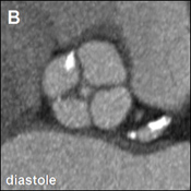
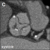
Double-oblique transverse multiplanar reconstruction during mid-diastole (70% of RR-interval) and during end-systole (300 ms past the R-peak) as well as TEE images are shown. A quadricuspid valve with four separate leaflets is seen. Leaflet margins were found to be thickened with mild to moderate calcifications. Diastolic reconstructions revealed a central zone of incomplete coaptation. By means of planimetry, this regurgitant orifice measured 0.12 cm, consistent with moderate aortic regurgitation seen on transesophageal echo (vena contracta 3 to 4mm). On systolic reconstructions, incomplete opening was observed (aortic valve area 0.8cm
2), consistent with severe aortic stenosis.
The correct answer is: C. Quadricuspid valves are usually associated with aortic valve regurgitation.
Quadricuspid aortic valve is a rare congenital malformation with an incidence between 0.003% and 0.013%, far less common than bicuspid aortic valves (incidence approx. 2%). It is frequently associated with aortic regurgitation, whereas valvular stenosis as shown in this case is very rare. (1)
References
- Janssens U, Klues HG, Hanrath P. Congenital quadricuspid aortic valve anomaly associated with hypertrophic non-obstructive cardiomyopathy: a case report and review of the literature. Heart 1997;78:83-7.