Poll: Mavacamten Use in Hypertrophic Cardiomyopathy
A 63-year-old woman presents to the office for a second opinion on chronic chest pain and heart failure management. She has a medical history of mixed connective tissue disease, obstructive sleep apnea, hepatic steatosis, and long-standing hypertension diagnosed in her twenties.
She reports several years of resting and exertional chest pain occurring up to five times weekly, shortness of breath with most daily activities, generalized weakness, and exercise intolerance. She has had minimal relief from diuretic or antianginal therapy, including long-acting nitrates, beta-blockers, calcium channel blockers, and ranolazine. Her symptoms have progressed in frequency and severity over the previous 6 months. Her current medications are metoprolol succinate 25 mg daily, losartan 50 mg daily, eplerenone 100 mg daily, ranolazine 500 mg every 12 hours, and furosemide 20 mg daily. A recent coronary angiogram had findings of normal epicardial coronary arteries.
She reports that her paternal grandfather died from a myocardial infarction (MI) at 42 years of age. Her son had a MI at 33 years of age. No other family members carry a history of heart disease.
Today, her examination demonstrates blood pressure 141/76 mm Hg, heart rate 62 bpm, and weight 90 kg (body mass index 36 kg/m2). Cardiac auscultation reveals a regular rhythm with no resting murmur and faint grade 1/6 systolic ejection murmur with the Valsalva maneuver. Her jugular venous pressure is normal, and she has no peripheral edema. Laboratory test findings are notable for N-terminal pro–B-type natriuretic peptide level 75 pg/mL; comprehensive metabolic panel and complete blood count values are within the reference ranges.
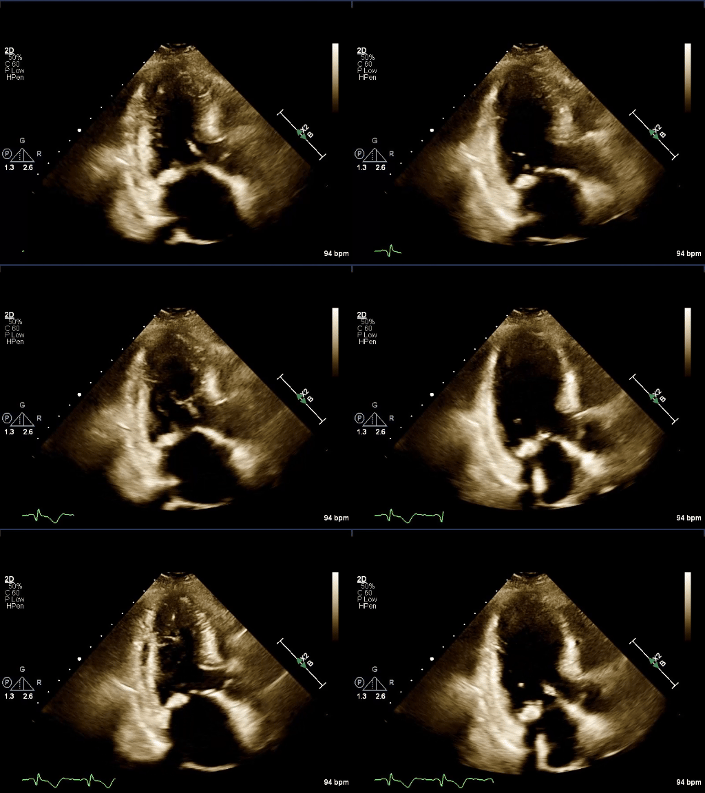
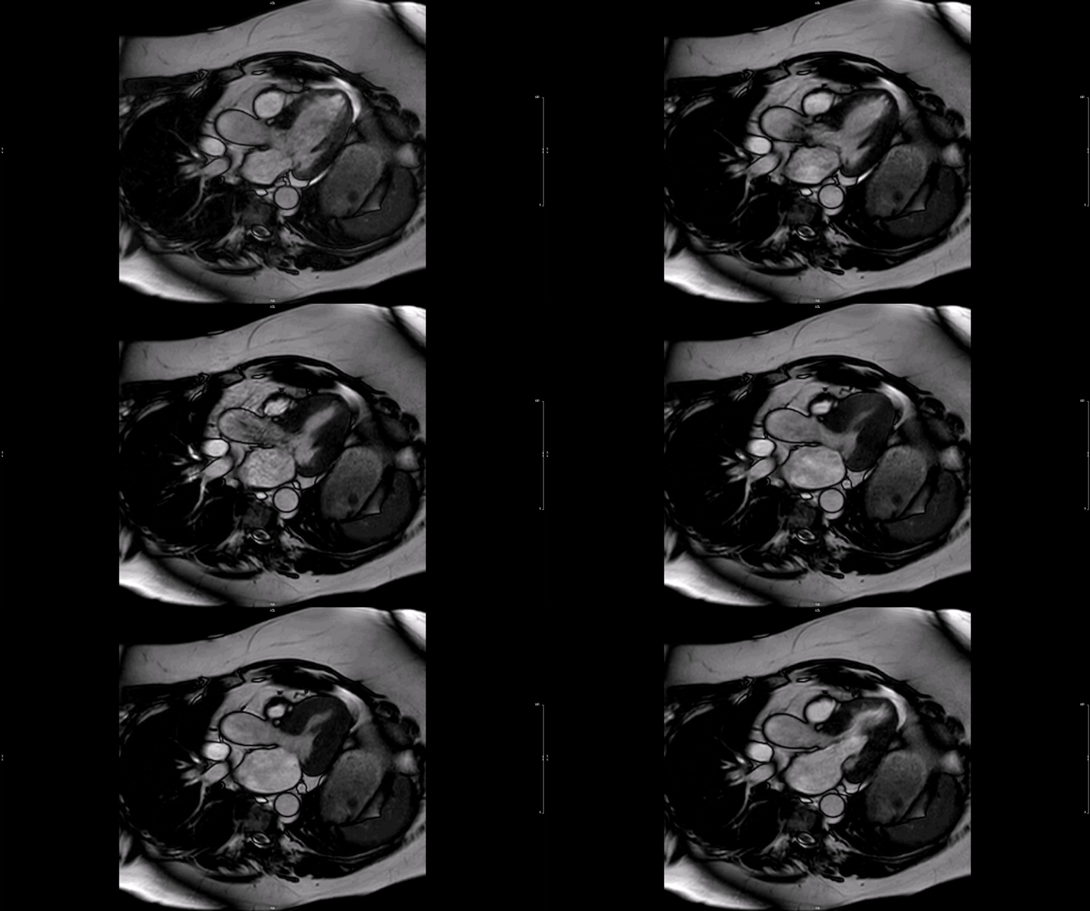
A transthoracic echocardiogram has findings of normal left ventricular (LV) function with estimated LV ejection fraction 60%. There is asymmetric LV hypertrophy with maximal septal thickness 15 mm at the inferoseptum. Midventricular flow acceleration and papillary muscle–septal contact is seen (Video 1). There is a dynamic midventricular gradient (10 mm Hg at rest, 59 mm Hg with the Valsalva maneuver). Cardiac magnetic resonance imaging findings include small LV size with hyperdynamic function, near-complete mid-to-apical systolic cavity obliteration, and prominent papillary muscle–septal contact (Video 2). No systolic anterior motion of the mitral valve is present.
Video 1

Video 2

Clinical Topics: Heart Failure and Cardiomyopathies, Noninvasive Imaging