Studies Offer New Insight Into LBBAP Mechanisms
Two studies recently published in JACC: Clinical Electrophysiology draw a sharp focus on modalities of left bundle branch area pacing (LBBAP), including left bundle branch pacing (LBBP) and left ventricular septal pacing (LVSP), as well as expand the current understanding of underlying physiological mechanisms.
An analysis from the prospective TREEBEARD study found that LBB pacing (LBBP) preserves left ventricular (LV) mechanical efficiency comparable to spontaneous LV activation in patients with preserved or moderately reduced LVEF, based on correlations with indices of myocardial work (MW).
Matteo Bertini, MD, PhD, et al., assessed MW among 155 study patients with preserved or moderately reduced LVEF (73 years old, 25% women) who underwent either LBBP (n=102) or LVSP (n=53).
Results showed no significant differences between the LBBP and LVSP groups for spontaneous QRS duration or for MW indices except for global work efficiency (GWE) which was 94.1 mm Hg% and 86.5 mm Hg%, respectively.
Additionally, there was a strong correlation between values recorded at spontaneous rhythm and those recorded at LBBP for all assessed MW metrics: global work index (GWI; r=0.881), global constructive work (GCW; r=0.742), global wasted work (GWW; r=0.758) and GWE (r=0.748) (p<0.001 for all).
However, in the LVSP group, there were only moderate correlations in GWI (r=0.535; p=0.004), GCW (r=0.587; p=0.009) and GWE (r=0.503; p=0.01), with no significant correlation with GWW (r=0.641; p=0.13).
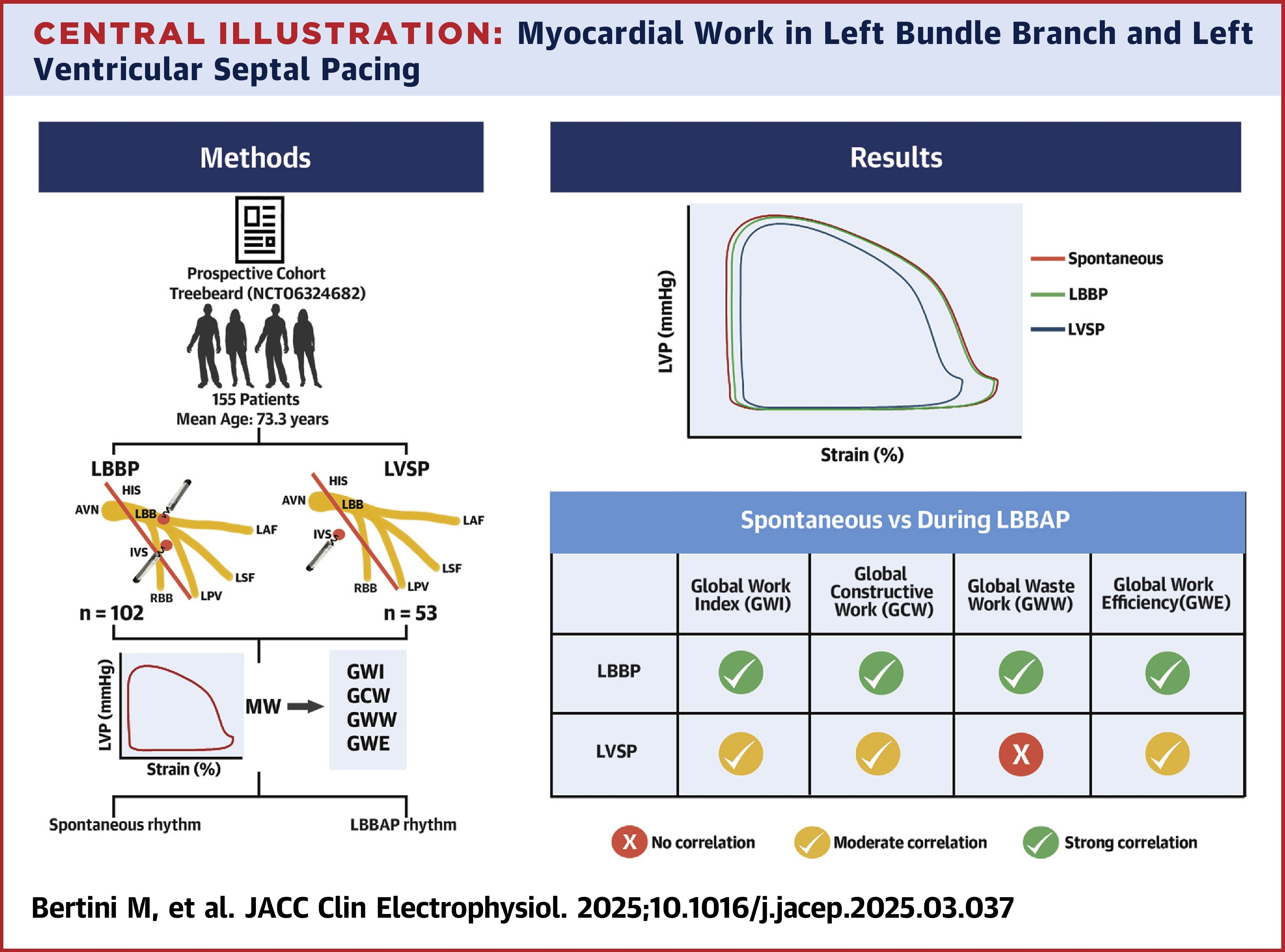
The authors note that LVSP is a "valuable option" in this patient population. Moreover, they write, "These findings underscore the overall advantage of pacing that engages directly the [left bundle], particularly when achieving LBBP for maximal energetic and physiological benefit."
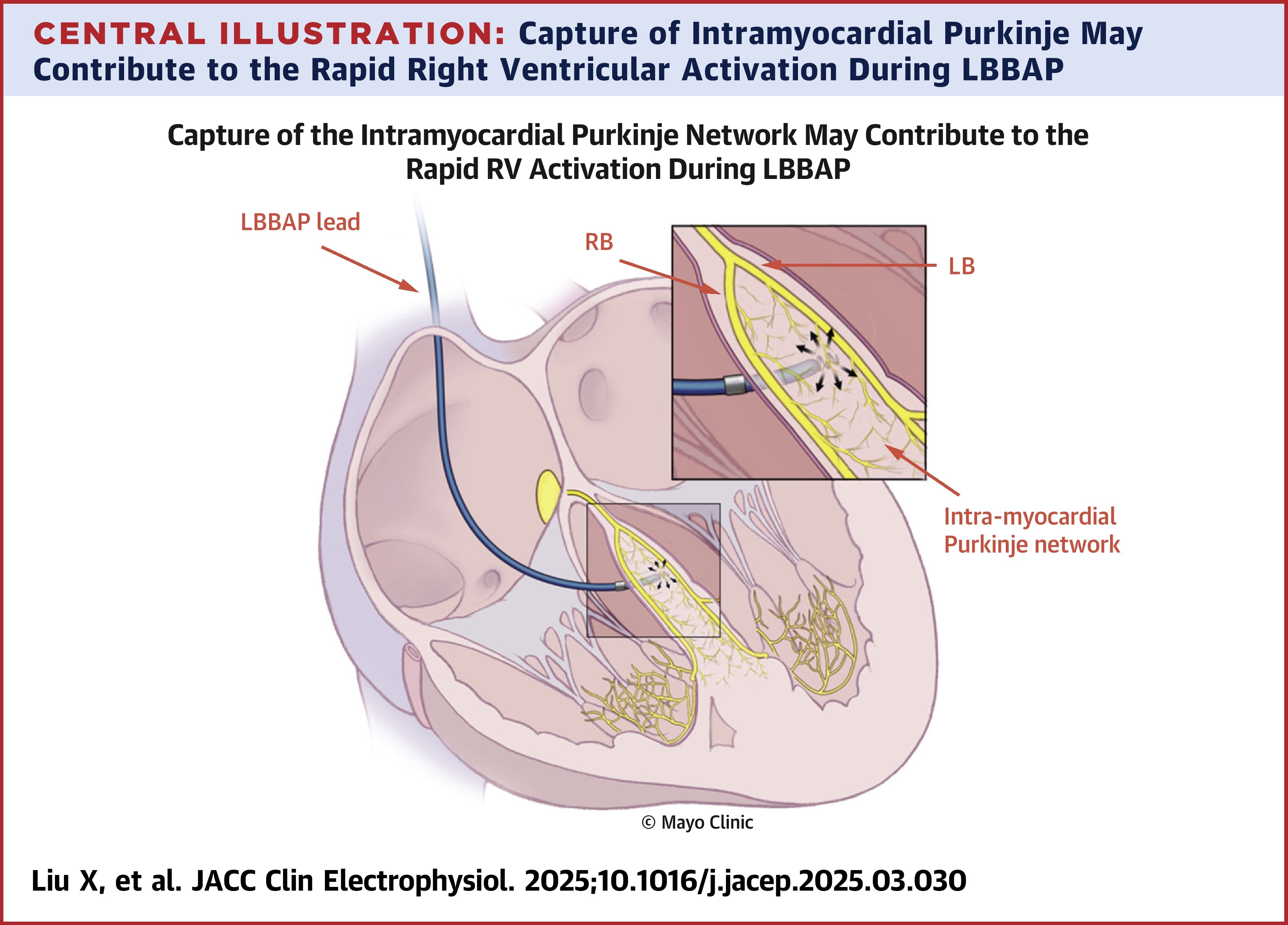
Another study builds on current knowledge of LBBAP mechanisms, potentially attributing previously unexplainable abnormal electrocardiogram and intracardiac recordings to the capture of the intramyocardial Purkinje network – contributing to rapid right ventricular (RV) activation.
Xiaoke Liu, MD, PhD, et al., evaluated 13 patients with surface electrocardiogram (ECG) or intracardiac recording features unexplained by selective or nonselective LBBAP and analyzed septal Purkinje fiber staining patterns in human cardiac tissue.
They uncovered five unusual ECG and intracardiac recording patterns unexplained by prevailing LBBAP theories: 1) alteration of right bunch branch block (RBBB) and LBBB as output-independent and -dependent, 2) variable recruitment of the left and right bundle systems, 3) correction of baseline RBBB at low outputs, 4) paced QRS axis and duration closely matching baseline narrow QRS interval in patients who underwent atrioventricular node ablation, and 5) intracardiac recordings demonstrating rapid, apparent nonphysiological activation of the RV septum.
Results from the analysis of the human cardiac tissue revealed dense Purkinje tissue deep inside the septal myocardium near the usual location of the LBBAP lead, "[challenging] the conventional understanding that the human conduction system and its arborizing Purkinje fibers are exclusively situated near the endocardium," according to the authors.
"We postulate that direct capture of this network connected to both left and right bundles branches followed by rapid propagation through the rest of the bundle systems to activate both ventricles could provide a unifying explanation for all the counterintuitive findings presented in this series," they conclude.
Citations:
- Bertini M, Vitali F, Malagù M, et al. Left Ventricular Mechanical Insights Into Left Bundle Branch Pacing and Left Ventricular Septal Pacing. JACC Clin Electrophysiol. Published online May 29, 2025. doi:10.1016/j.jacep.2025.03.037
- Liu X, Mulpuru SK, Behfar A, et al. Septal Intramyocardial Purkinje Network: A Potential New Mechanism Explaining Left Bundle Branch Area Pacing Physiology. JACC Clin Electrophysiol. Published online May 29, 2025. doi:10.1016/j.jacep.2025.03.030
Clinical Topics: Arrhythmias and Clinical EP, EP Basic Science
Keywords: Purkinje Fibers, Ventricular Septum
< Back to Listings