Editor's Note: This is the second half of a two-part Patient Case Quiz. Go to Part I.
Case B Presentation: A 32 year old female with no past medical history presents to your clinic for a second opinion regarding her chronic pericardial effusion. The effusion was discovered on an echocardiogram performed six years ago for palpitations which are no longer problematic. Over the past year, she has developed central chest pain that typically lasts for 1 minute and occurs five to ten times daily. At baseline, she is very active and recently underwent an exercise stress test (peaked at 13 METS). Her local physician follows the effusion with yearly echocardiograms (all demonstrating stability in size). She was prescribed a two week course of a non-steroidal anti-inflammatory agent but this has failed to improve her symptoms or reduce the size of the effusion.
Physical exam: Pleasant young female, appears anxious. Blood pressure 134/76, pulse 90, respirations 12. Body mass index 20 kg/meter-squared. Her JVP is 6 cm, lungs are clear with no gallop or murmur. Her PMI is non-displaced, skin is warm, and there is no lower extremity edema. Abdominal exam reveals no organomegaly.
Laboratory testing: Thyroid stimulating hormone normal; Sedimentation rate 5 mm/hr; C-reactive protein 0.3 mg/L; CK-MB negative, Troponin negative. ANA and Rheumatoid Factor were negative. Sodium 138, Potassium 3.8, Chloride 110, Bicarbonate 24, Blood urea nitrogen 7, Creatinine 0.62. She had normal liver enzymes and a normal complete blood count.
EKG: Normal sinus with premature atrial complexes. PR interval is 120 milliseconds, QRS duration is 72 milliseconds. Her corrected QT is 430 milliseconds.
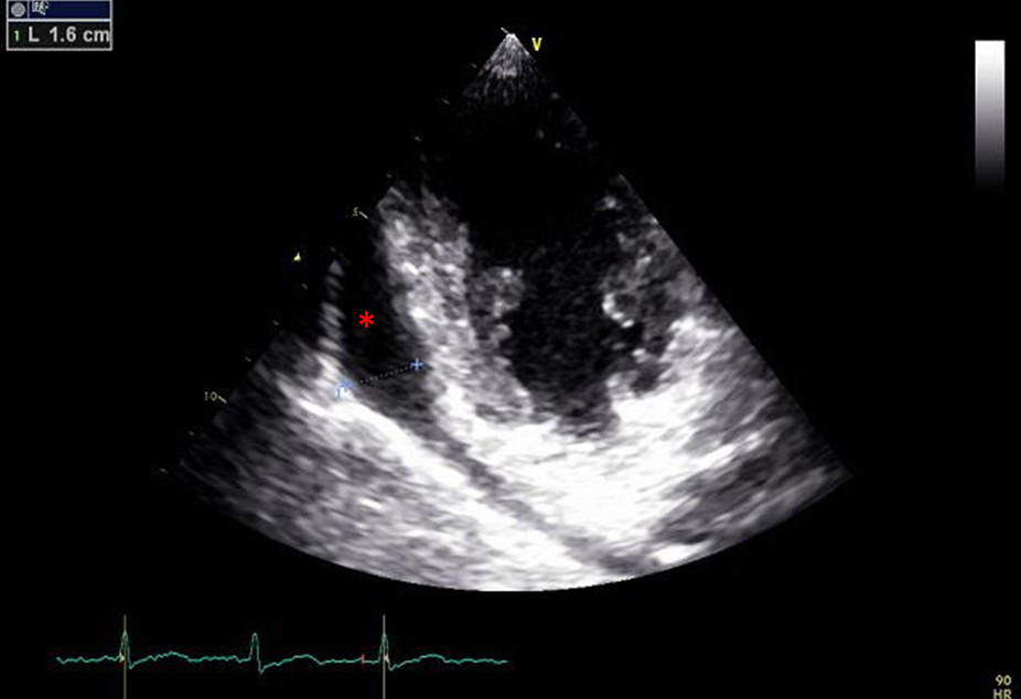
Echocardiogram: The left and right ventricles are normal in size and systolic function. Diastolic function is normal. The estimated right ventricular pressure is normal. The left and right atrial cavities are normal in size. There are no major valvular abnormalities. There is a moderately sized circumferential pericardial effusion measuring 16 mm posteriorly with no right atrial or ventricular diastolic inversion.
Cardiac MRI: No pericardial delayed enhancement.
Two-dimensional echocardiogram shows a moderate-sized circumferential pericardial effusion (red asterisk). The effusion measures 16 mm.
Video 1: Short Axis
Video 2: Long Axis
How would you manage the chronic idiopathic pericardial effusion?
Show Answer
The correct answer is: D. Serial follow-up echocardiogram
The patient in this case is asymptomatic with negative inflammatory markers, but has a moderately-sized circumferential effusion. She is at lower risk for progression to tamponade given the size of her effusion and can be followed with serial echocardiograms. Further trial of anti-inflammatory agents is not necessary in this case since there was no evidence of active inflammation on MRI.
Pericardial effusions that persist for more than three months are by convention chronic. As a general approach, all chronic effusions should be evaluated for size, hemodynamic effect (e.g. right sided collapse), ability to produce symptoms, and etiology. Although the etiology of chronic effusions can often be elusive, a thorough workup should be conducted since a specific cause dictates treatment and prognosis. This can include age-appropriate cancer screening, assessing for thyroid disorders, and searching for infectious, inflammatory, and rheumatologic diseases. The diagnostic yield for a pericardial tap is very low (approximately 7%) and thus should not be routinely performed unless an infectious or malignant process is highly suspected.1
The natural history of chronic idiopathic pericardial effusions and the propensity to develop tamponade physiology is not well studied. Management is largely empirical. Conflicting reports exist in the literature from few studies with limited number of patients. Most suggest that large (>20 mm) idiopathic effusions are well tolerated for long periods of time; however, progression to clinical tamponade is unpredictable and can be seen in up to 30% of cases.2 The key management question centers around which patients should be referred for a tap or surgical window over conservative management since an unnecessarily invasive approach may trigger iatrogenic pericarditis. Possible predictors for tamponade include size (>20 mm), recurrent acute pericarditis, chest wall trauma, hypovolemia, right sided collapse on echocardiogram, or the presence of a paroxysmal tachyarrhythmia.3,4 The European Society of Cardiology has published a triage strategy for urgent management of cardiac tamponade that could potentially be applied to large chronic idiopathic effusions.5 In their scoring system (which is not validated), etiology, symptoms, and imaging are all scored. Symptoms of orthopnea and a rapid progression of overall symptoms are weighed the highest in the clinical presentation category. In the imaging category, circumferential effusion > 20 mm, right ventricular collapse, less than 50% inspiratory collapse of an IVC that is > 25 mm, and left atrial collapse are given the greatest weight.
Medical therapy with anti-inflammatory agents (NSAID, colchicine, steroids) can be tried if the C-reactive protein or sedimentation rate is elevated or if there is evidence of delayed enhancement on cardiac MRI. However, these agents are generally not efficacious if inflammatory markers are normal or if the cardiac MRI is not suggestive of active pericardial inflammation.4
References
Merce J, Sagrista-Sauleda J, Permanyer-Miralda G, Soler-Soler J. Should pericardial drainage be performed routinely in patients who have a large pericardial effusion without tamponade? The American journal of medicine 1998;105:106-9.
Sagrista-Sauleda J, Angel J, Permanyer-Miralda G, Soler-Soler J. Long-term follow-up of idiopathic chronic pericardial effusion. The New England journal of medicine 1999;341:2054-9.
Imazio M, Spodick DH, Brucato A, Trinchero R, Adler Y. Controversial issues in the management of pericardial diseases. Circulation 2010;121:916-28.
Imazio M, Adler Y. Management of pericardial effusion. European heart journal 2013;34:1186-97.
Ristic AD, Imazio M, Adler Y et al. Triage strategy for urgent management of cardiac tamponade: a position statement of the European Society of Cardiology Working Group on Myocardial and Pericardial Diseases. European heart journal 2014;35:2279-84.