Innovation in CV Imaging
Technology provides an excellent opportunity to diagnose the vague cardiovascular disease. Especially in recent years, several technological advances in diagnosis and therapy have significantly changed clinical practice.
Cardiology has become increasingly interventional as many new capabilities have diffused from tertiary medical centers to the community. These developments have contributed to observed reductions in death rates from heart disease and improvements in the quality of life.
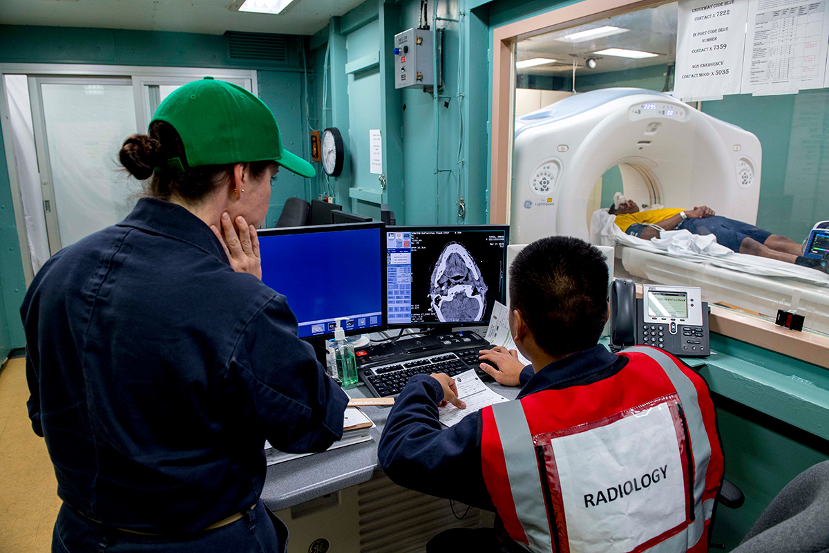
Numerous techniques are available for producing images of the heart that provide valuable information for guiding diagnosis, patient assessment and therapeutic intervention, including cardiac imaging techniques, 3D echocardiography, computed tomography with measurement of coronary fractional flow reserve, etc.
These techniques have become a central pillar in the diagnosis of a variety of heart conditions other than coronary disease, such as valve disease, septal defects and wall motion abnormalities.
Through monitoring, these techniques can scrutinize the results of examination and telemedicine, as well as analyze the present and future of cardiac tissue engineering and organogenesis.
In the coming years, we are likely to see continuing improvements in cardiovascular imaging accuracy and the breadth of clinical information it generates, and consequently, echocardiography may pose a substantial competitive threat to diagnostic nuclear cardiology.
The recent history of cardiovascular imaging illustrates the flexibility and power of this technology. The M-mode machines used in early clinical diagnoses could look only along a single axis, providing what was sometimes called an "icepick" view.
An ultrasonic signal would be transmitted along this axis and reflected off any anatomical structures it encountered.
As these structures moved, differences in the length of the travel path created corresponding differences in the time between emission and detection of the reflection. Originally, these machines were limited to the diagnosis of valvular disorders.
The later addition of scanning and more advanced signal processing capabilities enabled clinicians to generate 2D images of the heart. With this development, users of echocardiography could examine large-scale cardiac anatomy.
The technology quickly evolved from still pictures to real-time moving images. Moving pictures provided valuable information on cardiac function. Changes in volume over the course of the beat cycle provided a basis for assessing the heart's pumping ability.
By revealing wall motion abnormalities, real-time imaging also provided indirect information on the presence of scar tissue and/or ischemia.
The development of pulse Doppler allowed echocardiography to use changes in frequency caused by the motion of the blood in the heart to generate quantitative information about velocities.
Other developments have increased the accuracy and usefulness of this technology. For instance, the development of reliable probes that could be inserted into the esophagus improved the resolution of these images by permitting clinicians to view the heart without the distortions caused by the passage of signals through fat, bone, and lung tissue.
The more recent development of acoustic quantification using techniques to detect and monitor the boundary between the inner edge of the myocardium and blood pool that it contains has provided automated real-time measurements of ventricular function information, which has been shown to have major prognostic significance.
A number of further efforts to increase the accuracy of echocardiography are currently underway. These techniques can determine the amount of blood supplied to different regions of the heart walls.
On the other hand, investigation involves the development of miniature probes that can be inserted into the heart through the coronary vasculature. One clinician has predicted that intraluminal ultrasound imaging using these probes will eventually displace angiography as the "gold standard" for detecting and localizing coronary artery disease.
Other effort is attempting to use subtle aspects of the ultrasound signal to characterize the tissues through which the signal has passed. Investigators have attempted to use this technique to identify tumors and distinguish between scar tissue and ischemic myocardium.
Successful achievement of these goals would also firmly position echocardiography as a direct competitor of nuclear cardiology.
An innovation in cardiovascular imaging could be clinically useful.
Additionally, new software in advanced echocardiography are using detailed assessments of movement in both left and right ventricles' segments. The computer- assisted images and SPECT camera can be helpful to innovative new aspects of this technique in the near future.
The eventual significance of an innovation is not usually apparent at the time of concept development. However, our goal is emphasis on creation in imaging, and identification of this would play a significant role in the efforts to develop the best future for imaging and echocardiography.

This article was authored by Azin Alizadehasl, MD, FACC, professor of cardiology and echocardiography, and head of the Cardio-Oncology Department and Research Center at the Rajaie Cardiovascular Medical and Research Center in Tehran, Iran.