A 26 year-old male with a history of viral myopericarditis one year prior presented to cardiology clinic for a follow-up visit. The patient was a recreational athlete who had been playing full-court basketball with a league five to six times a week for many years. One year prior to this index presentation, he had presented to the emergency department with three days of mid-sternal chest pain relieved by leaning forward. His symptoms began one week after an upper respiratory tract infection. His electrocardiogram (ECG) was notable for left ventricular hypertrophy with early repolarization, and his initial troponin T was 0.406 ng/mL. An echocardiogram was unremarkable. His troponin downtrended to 0.380 ng/mL the following day. He was seen by cardiology who felt his presentation was consistent with mild viral myopericarditis, and recommended activity restriction along with close follow-up. He was discharged on a regimen of high-dose ibuprofen for two weeks, along with daily colchicine 0.6mg for three months. His symptoms quickly resolved. Six months later, after labwork confirmed normalization of inflammatory biomarkers, he underwent a maximal-exercise ECG treadmill stress test which was unremarkable. He was permitted to return to playing moderate to high intensity recreational basketball at that time.
Three months later, he presented to a different emergency department after a 10-day history of an upper respiratory tract infection. He described a subacute onset of chest pain and dyspnea during that time, similar to his symptoms nine months prior, but more intense in nature. An ECG was reportedly unremarkable, however an initial troponin I was markedly elevated at 20 ng/mL. He was admitted and underwent further testing with an echocardiogram and coronary computed tomography angiography (CTA), both of which were normal. A basic rheumatologic workup was also unrevealing. His chest discomfort improved with the initiation of colchicine and high dose ibuprofen, and his troponin I downtrended to 15 ng/mL on the day of discharge. He was again advised to restrict his physical activity to only low exertion. Two weeks later he presented to clinic for follow-up evaluation.
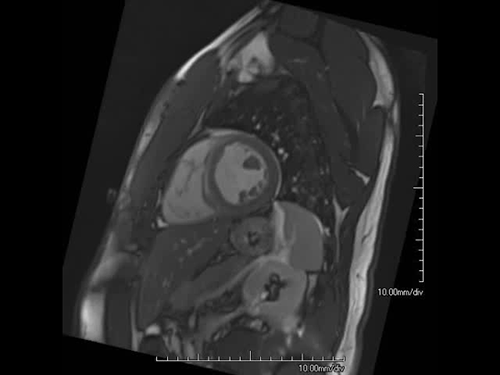
At this index visit, he reported complete resolution of his symptoms and adherence to low activity restriction. Vital signs were within normal limits. Physical exam revealed clear lungs, a normal jugular venous pulsation, and a regular cardiac rhythm, without any murmurs, rubs or gallops. His extremities were warm without any edema. An ECG again showed left ventricular hypertrophy without additional abnormal findings. Labwork at this visit showed normal chemistries and a complete blood count, as well as normal inflammatory markers. Cardiac magnetic resonance imaging (MRI) was obtained; demonstrative cine and sequential delayed gadolinium enhancement (DGE) images are shown in Videos 1 and 2.
Video 1 – Cine
Video 1 – Cine
Video 2 – Axial Stack Sequence
Based on the MRI findings, what is the next best step in management?
Show Answer
The correct answer is: D. All of the above.
The cardiac MRI revealed a mildly dilated left ventricle (LV) with mildly increased anteroseptal and inferoseptal wall thickness. There was mild hypokinesis of the inferoseptum, inferior, and inferolateral walls from mid-wall to apex. The LV ejection fraction was mildly decreased at 45%. Significant mid- to subepicardial DGE was seen in the septal and inferior walls from base to mid-wall in a spiral pattern consistent with a history of myocarditis. The right ventricle was also noted to be mildly increased in size, with mildly reduced systolic function.
Current US guidelines recommend that athletes with a probable or definite diagnosis of recent myocarditis be restricted from participating in competitive sports while inflammation is present. Resolution of active inflammation is variable and can take months. There is no single test to confirm resolution of inflammation, however, guidelines suggest it is reasonable to return to play as long as there is no biomarker evidence of inflammation and no concern for clinically relevant arrhythmias. As such, it is advised that athletes undergo clinical testing with an exercise ECG, echocardiogram, and rhythm monitoring no less than three to six months after the initial illness prior to lifting restriction on moderate to vigorous physical activity.1
The pathogenesis of viral myocarditis is thought to be composed of three confluent phases: acute viral injury, followed by host immunologic responses, and finally, recovery or a transition to scar and dilated cardiomyopathy.1,2 It is thought that cardiomyopathy associated with acute myocarditis often resolves over 6 to 12 months, however, individual responses and courses are highly variable. Athletes in whom evidence of active inflammation has resolved may still have a risk of arrhythmias related to myocardial scar. Studies have shown that the presence of DGE confers a worse prognosis among those with prior myocarditis and residual scar.3
While the role of follow-up MRI to evaluate for resolution of DGE remains unclear, the reintroduction intense physical activity depends on the severity of LV dysfunction and the extent of recovery. In general, patients with myocarditis and acute LV dysfunction should be treated according to current heart failure guidelines.2 As such, initiation of a beta blocker and ACE-inhibitor as tolerated, along with continuation of anti-inflammatory therapy would be advised in this situation. It would also be acceptable to repeat a cardiac MRI after 3-6 months in this case to assess for burden of DGE as well as residual LV dysfunction, in addition to guideline-recommended testing. Following this, a shared-decision making approach to safe return to activity should be conducted with the patient.
The patient was initiated on a low-dose ACE-inhibitor and beta-blocker as tolerated, along with 3 months of colchicine. A 2-week rhythm monitor was obtained 3 months after his visit without evidence of clinically significant arrhythmias. He was subsequently lost to follow-up due to loss of employment and consequently his health insurance. Now over a year later, he is actively employed and has returned for follow-up. He self-discontinued his colchicine in the interim, but reports continued adherence to activity restriction as well as to his beta-blocker and ACE-inhibitor without recurrence of symptoms. He remains interested in returning to play recreational basketball. While not mandated by guidelines, a cardiac MRI has been ordered to re-assess LV function and residual DGE prior to maximal exercise stress testing in order to inform future conversations about safe return to play.
References
Maron BJ, Zipes DP, Kovacs RJ. Eligibility and disqualification recommendations for competitive athletes with cardiovascular abnormalities: preamble, principles, and general considerations: a scientific statement from the American Heart Association and American College of Cardiology. J Am Coll Cardiol 2015;66:2343-49.
Cooper LT Jr. Myocarditis. N Engl J Med 2009;360:1526-38.