When and How to Test for Liver Disease in the Fontan Population
The Fontan Circulation
Since the Fontan operation was first proposed as a corrective procedure for tricuspid atresia, it has evolved to become the definitive palliative surgery for individuals born with single ventricle physiology or for those in whom a biventricular repair is not feasible. As a result, thousands of individuals with complex congenital heart disease have reached adulthood who otherwise would not have survived.1
By redirecting the systemic venous return into the pulmonary circulation, albeit without an intervening ventricle, a successful Fontan procedure results in normalization of systemic oxygen saturation and ventricular volume load. At the same time, the lack of a subpulmonary ventricle compromises preload to the systemic ventricle, and the arrangement of having the systemic and pulmonary vascular beds lie in series increases ventricular afterload. These adverse hemodynamic consequences conspire to depress cardiac output at rest and to limit augmentation of cardiac output during stress.2 In addition, the “passive” pulmonary blood flow in the Fontan circulation requires chronically elevated central venous pressures compared to those in individuals with two-ventricle physiology.
This altered physiology of passive flow through the pulmonary arterial bed often results in failure of the Fontan circulation as the patient ages. Comorbidities include arrhythmia, thromboembolic events, protein-losing enteropathy, plastic bronchitis and heart failure.3 Liver disease related to the Fontan circulation has garnered widespread attention among cardiologists only recently, but it is increasingly felt to be an important contributor to morbidity years after Fontan surgery.4
The Liver in Heart Disease
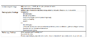
The relationship between congestive heart failure and liver disease has been recognized since the early 19th century when heart disease was noted to be associated with hepatic dysfunction and cirrhosis.5 However, the impact of liver disease on the aging cohort of Fontan patients may be particularly pronounced due the chronicity of the Fontan circulatory arrangement. The elevated central venous pressure in patients with Fontan circulation is most commonly implicated in the pathogenesis of Fontan liver disease.6 However, other potential mechanisms for Fontan-related liver disease that deserve consideration include pre-Fontan hypoxemia,7 perioperative liver injury8, 9 and altered hepatic blood flow.10, 11 Potential laboratory, imaging and histopathologic findings are summarized in Table 1.
Imaging abnormalities
In patients who have been subjected to very basic or no screening of their liver health, it is often abdominal imaging for other indications that first calls attention to the liver. Ultrasound imaging is often characterized by hepatomegaly with hepatic vein and suprahepatic inferior vena cava dilation. In more advanced liver disease, surface nodularity, increased parenchymal echogenicity, and an irregular parenchymal echotexture may be noted. CT or MR imaging often shows hepatomegaly, splenomegaly and collateral vessels, including intra- and extrahepatic venovenous collaterals, large gastroesophageal varices, and portopulmonary venous collaterals. Abnormal contrast enhancement on CT, zonal or reticular in distribution, is typical. Hypervascular nodules may be seen and can be confused with tumors. They are thought to represent arterialization of the liver secondary to decreased portal vein inflow from increased central pressures.12, 13
Laboratory abnormalities
Liver disease can be easily overlooked in Fontan patients because abnormalities in routine liver tests tend to be mild. The most common abnormal finding is an elevation in gamma-glutamyl transferase (GGT), noted in 40-60% of patients in the outpatient setting.14-18 However, this test is not usually included in the commonly-ordered “hepatic function panel” or “comprehensive metabolic panel” offered by many labs. Elevated serum levels of aminotransferases (i.e., alanine aminotransferase and aspartate aminotransferase), markers of liver injury, are reported in about one-third of Fontan patients. This reflects the fairly modest hepatocyte injury seen in most cases. In decompensated patients with poor cardiac output, however, aminotransferase levels may become markedly elevated as a result of hepatic ischemia.
The serum bilirubin is elevated in 25-40% of patients and tends to be mild and mostly unconjugated. The cause of hyperbilirubinemia in Fontan patients is not fully understood, but contributing factors may include hepatocellular dysfunction, hemolysis, canalicular obstruction from distended hepatic veins, and medications.
Total protein and albumin levels, indicators of liver synthetic function, are decreased in only 5-10% of patients seen in the outpatient setting. Prothrombin time (PT) is abnormal in as many as 65-79% of older cohorts late after Fontan. This is generally related to deficiencies in liver-derived coagulation factors including proteins C and S; factors II, V, VII, IX, and X; antithrombin III; and plasminogen.14-16, 18
Histologic abnormalities
In the more common presentation of liver disease in the setting of congestive heart failure, the characteristic gross appearance is frequently described as “nutmeg liver,” with areas of sinusoidal congestion and sometimes hemorrhagic necrosis appearing reddish-brown on a yellowish background of normal or sometimes fatty liver parenchyma. The typical histologic findings include sinusoidal dilation, hepatocyte atrophy, and varying degrees of centrilobular fibrosis ranging from mild perisinusoidal collagen deposition to “reverse lobulation,” in which portal tracts are seen in the center of a “lobule” formed by broad, fibrous bands connecting central vein to central vein.6, 19
Compared with the hepatic changes typically reported in congestive heart failure (CHF), the abnormalities seen in Fontan patients are characterized as being more severe: the degree of sinusoidal dilatation is more extreme, and the extent of fibrosis and fibrous septum formation is more severe than that generally described in long-standing heart failure.
“But Is Any of This Clinically Relevant?”
Is the presence of liver disease a marker of worsening Fontan status or a clinically important phenomenon? The implications of liver abnormalities have not been studied rigorously in the Fontan population. In non-congenital heart disease populations, cirrhosis predicts poor clinical outcomes after cardiac surgery,20-23 ventricular assist device (VAD) implantation and cardiac transplant,24, 25 and based on this, advanced hepatic fibrosis often even adversely influences a patient’s candidacy for cardiac transplantation.
When liver function was assessed in a cohort of Fontan patients by measuring indocyanine green clearance, disappearance and retention rates were significantly abnormal and similar to patients with viral cirrhosis, suggesting that laboratory and imaging abnormalities may coincide with limitation of liver function.26 Clinical consequences attributable to liver disease such as ascites and hepatic encephalopathy are not rare in the Fontan population and single ventricle physiology is associated with non-alcoholic cirrhosis, congestive liver disease, longer hospital stay and higher cost.27 When taken together, these data suggest that liver disease is clinically relevant, can be assessed by laboratory and imaging studies, and are reflective of true liver dysfunction which confers additional morbidity to the Fontan patient.
Particularly concerning is the number of case reports of patients with Fontan circulation who have developed hepatocellular carcinoma on the background of chronic passive congestion and fibrosis, even in the absence of frank cirrhosis.28-31 Cirrhosis is known to be associated with an increased risk of hepatocellular carcinoma, but congestive hepatopathy has not been considered to be a high-risk condition. Current guidelines suggest that screening for HCC be performed for all forms of cirrhosis.32
How and When Should We Test for Liver Disease?
Given the indolent, yet progressive, nature of liver disease in this population, patients who have already developed symptoms are likely to have more advanced disease, and the window of opportunity for effective intervention may be long past. Liver disease appears endemic in the older Fontan population and contributes to poor outcomes as these patients age. There is near universal fibrosis among patients included in biopsy studies, raising the concern that we are not looking for liver disease until it is too late. Therefore, we advocate for regular surveillance of liver health in all adolescent and adult Fontan patients at their routine follow-up visits.
Traditional serum markers for liver disease are insensitive and nonspecific, biomarkers have not been validated in the Fontan population, hepatic CT has not been established to correlate with histopathology, and diffusion weighted MRI may quantify fibrosis but has not been studied in the Fontan population.12, 13, 31, 33, 34
The gold standard for assessment of liver disease is liver biopsy. However, it is an invasive procedure, carries potential for bleeding with elevated venous pressures, may not be feasible in patients with significant ascites, and there is potential for sampling error. In addition, clinicians may be unsure of how to use the data obtained from the biopsy.
Which studies are appropriate to do routinely and when to proceed to more involved testing remains an open question. In the absence of known liver disease, we feel it is reasonable to check standard hepatic labs (i.e., AST, ALT, GGT, albumin, total protein, and total bilirubin), a complete blood count (declining platelet counts may indicate portal hypertension) and a prothrombin time once a year. Imaging can be performed with either CT or MRI every three to four years. The need for further testing such as liver biopsy would then be dictated based on these findings or on physical findings suggestive of advanced liver disease such as hepatomegaly, splenomegaly, gynecomastia, vascular ectasias, or jaundice. Biopsy can be performed through the transjugular route with a lower risk of bleeding and can be performed concurrently with cardiac catheterization. However, problems with adequate sample size can limit the diagnostic utility of transjugular biopsy, and a percutaneous biopsy is often necessary. Some institutions that see a large number of Fontan patients now endorse routine cardiac catheterization and liver biopsy at a pre-specified interval after Fontan surgery.35 In addition, the field of ACHD is also moving toward the development of reliable, less invasive alternatives for surveillance as we catalog and assess the clinical impact of liver disease in the Fontan population.
Screening for Hepatocellular Carcinoma
Also important to consider is screening for hepatocellular carcinoma. Modern imaging techniques can effectively identify tumors smaller than 2 cm which confer a better prognosis than larger lesions, and screening has been demonstrated to be cost effective in populations at increased risk. Because risk factors for the development of hepatocellular carcinoma specific to the Fontan population are not known, we recommend that any patient with histological evidence of advanced fibrosis (stage III-IV/IV using the Knodell scoring system36) undergo routine surveillance.
| TABLE 1: Potential laboratory, imaging and histopathologic findings in Fontan patients with liver disease. |
 |
Traditionally, practitioners have used a combination of serum alpha-fetoprotein and ultrasonography, which increases detection rates but also increases cost and false-positive rates. As a result, the 2010 American Association for the Study of Liver Diseases (AASLD) guidelines on the management of hepatocellular carcinoma recommend only ultrasonography at six- to 12-month intervals.32 However, while ultrasonograms may be adequate for screening cirrhotics in general, we believe that non-ultrasound imaging modalities should be the preferred option in our patient population due to the high incidence of non-malignant vascular lesions in Fontan patients. Our experience has been that annual gadolinium-enhanced MRI, once advanced hepatic fibrosis has been established, is an ideal screening test. While CT is an adequate alternative for patients with contraindication to MRI, concerns about frequent radiation exposure mandated by this yearly screening protocol relegate it to second-line status.
Conclusions
The approach to liver health and to the systemic effects of hepatic dysfunction in single ventricle patients palliated with Fontan surgery presents a paradigm for the multidisciplinary, collaborative care team model that must be developed in order to ensure optimal care of our increasingly complex population of adolescents and adults with congenital heart disease. As our understanding of liver disease in the Fontan patient improves, recommendations for screening and management are likely to evolve. Cardiologists caring for patients with Fontan physiology should forge partnerships with subspecialty colleagues, including a hepatologist, within the context of a multidisciplinary “single ventricle survivorship team” invested in assessing and improving the care of this unique patient population.
References
- Rogers LS, Glatz AC, Ravishankar C, et al. 18 years of the Fontan operation at a single institution: results from 771 consecutive patients. J Am Coll Cardiol 2012;60:1018-25.
- Senzaki H, Masutani S, Kobayashi J, et al. Ventricular afterload and ventricular work in fontan circulation: comparison with normal two-ventricle circulation and single-ventricle circulation with blalock-taussig shunts. Circulation 2002;105:2885-92.
- Khairy P, Fernandes SM, Mayer JE, Jr., et al. Long-term survival, modes of death, and predictors of mortality in patients with Fontan surgery. Circulation 2008;117:85-92.
- Wu FM, Ukomadu C, Odze RD, Valente AM, Mayer JE, Jr., Earing MG. Liver disease in the patient with Fontan circulation. Congenit Heart Dis 2011;6:190-201.
- Rokitansky C, Swaine WE. A Manual Of Pathological Anatomy: Blanchard & Lea; 1855.
- Kendall TJ, Stedman B, Hacking N, et al. Hepatic fibrosis and cirrhosis in the Fontan circulation: a detailed morphological study. J Clin Pathol 2008;61:504-8.
- Ghosh ML, Emery JL. Hypoxia and asymmetrical fibrosis of the liver in children. Gut 1973;14:209-12.
- Jenkins JG, Lynn AM, Wood AE, Trusler GA, Barker GA. Acute hepatic failure following cardiac operation in children. J Thorac Cardiovasc Surg 1982;84:865-71.
- Matsuda H, Covino E, Hirose H, et al. Acute liver dysfunction after modified Fontan operation for complex cardiac lesions. Analysis of the contributing factors and its relation to the early prognosis. J Thorac Cardiovasc Surg 1988;96:219-26.
- Camposilvan S, Milanesi O, Stellin G, Pettenazzo A, Zancan L, D'Antiga L. Liver and cardiac function in the long term after Fontan operation. Ann Thorac Surg 2008;86:177-82.
- Narkewicz MR, Sondheimer HM, Ziegler JW, et al. Hepatic dysfunction following the Fontan procedure. J Pediatr Gastroenterol Nutr 2003;36:352-7.
- Bryant T, Ahmad Z, Millward-Sadler H, et al. Arterialised hepatic nodules in the Fontan circulation: Hepatico-cardiac interactions. Int J Cardiol 2010.
- Kiesewetter CH, Sheron N, Vettukattill JJ, et al. Hepatic changes in the failing Fontan circulation. Heart 2007;93:579-84.
- Cromme-Dijkhuis AH, Hess J, Hahlen K, et al. Specific sequelae after Fontan operation at mid- and long-term follow-up. Arrhythmia, liver dysfunction, and coagulation disorders. J Thorac Cardiovasc Surg 1993;106:1126-32.
- Nevens F, Lijnen P, VanBilloen H, Fevery J. The effect of long-term treatment with spironolactone on variceal pressure in patients with portal hypertension without ascites. Hepatology 1996;23:1047-52.
- Ozaki K, Kodama M, Yamashita F, et al. Esophageal varices without portosystemic venous pressure gradient in a patient with post-pericardiotomy constrictive pericarditis: a case report. J Cardiol 2005;45:161-4.
- Tomita H, Yamada O, Ohuchi H, et al. Coagulation profile, hepatic function, and hemodynamics following Fontan-type operations. Cardiol Young 2001;11:62-6.
- van Nieuwenhuizen RC, Peters M, Lubbers LJ, Trip MD, Tijssen JG, Mulder BJ. Abnormalities in liver function and coagulation profile following the Fontan procedure. Heart 1999;82:40-6.
- Sherlock S. The liver in heart failure; relation of anatomical, functional, and circulatory changes. Br Heart J 1951;13:273-93.
- Bizouarn P, Ausseur A, Desseigne P, et al. Early and late outcome after elective cardiac surgery in patients with cirrhosis. Ann Thorac Surg 1999;67:1334-8.
- Hayashida N, Shoujima T, Teshima H, et al. Clinical outcome after cardiac operations in patients with cirrhosis. Ann Thorac Surg 2004;77:500-5.
- Klemperer JD, Ko W, Krieger KH, et al. Cardiac operations in patients with cirrhosis. Ann Thorac Surg 1998;65:85-7.
- Lin CH, Lin FY, Wang SS, Yu HY, Hsu RB. Cardiac surgery in patients with liver cirrhosis. Ann Thorac Surg 2005;79:1551-4.
- Hsu RB, Lin FY, Chou NK, Ko WJ, Chi NH, Wang SS. Heart transplantation in patients with extreme right ventricular failure. Eur J Cardiothorac Surg 2007;32:457-61.
- Reinhartz O, Farrar DJ, Hershon JH, Avery GJ, Jr., Haeusslein EA, Hill JD. Importance of preoperative liver function as a predictor of survival in patients supported with Thoratec ventricular assist devices as a bridge to transplantation. J Thorac Cardiovasc Surg 1998;116:633-40.
- Guha IN, Bokhandi S, Ahmad Z, et al. Structural and functional uncoupling of liver performance in the Fontan circulation. Int J Cardiol 2011.
- Krieger EV, Moko LE, Wu F, et al. Single ventricle anatomy is associated with increased frequency of nonalcoholic cirrhosis. Int J Cardiol 2012.
- Ewe SH, Tan JL. Hepatotocellular Carcinoma—A Rare Complication Post Fontan Operation. Congenital Heart Disease 2009;4:103-6.
- Ghaferi AA, Hutchins GM. Progression of liver pathology in patients undergoing the Fontan procedure: Chronic passive congestion, cardiac cirrhosis, hepatic adenoma, and hepatocellular carcinoma. J Thorac Cardiovasc Surg 2005;129:1348-52.
- Saliba T, Dorkhom S, O'Reilly EM, Ludwig E, Gansukh B, Abou-Alfa GK. Hepatocellular carcinoma in two patients with cardiac cirrhosis. Eur J Gastroenterol Hepatol 2010;22:889-91.
- Wallihan DB, Podberesky DJ. Hepatic pathology after Fontan palliation: spectrum of imaging findings. Pediatr Radiol 2012.
- Bruix J, Sherman M. Management of hepatocellular carcinoma: an update. Hepatology 2011;53:1020-2.
- Baek JS, Bae EJ, Ko JS, et al. Late hepatic complications after Fontan operation; non-invasive markers of hepatic fibrosis and risk factors. Heart 2010;96:1750-5.
- Ginde S, Hohenwalter MD, Foley WD, et al. Noninvasive assessment of liver fibrosis in adult patients following the Fontan procedure. Congenit Heart Dis 2012;7:235-42.
- Rychik J, Veldtman G, Rand E, et al. The precarious state of the liver after a Fontan operation: summary of a multidisciplinary symposium. Pediatr Cardiol 2012;33:1001-12.
- Knodell RG, Ishak KG, Black WC, et al. Formulation and application of a numerical scoring system for assessing histological activity in asymptomatic chronic active hepatitis. Hepatology 1981;1:431-5.
Clinical Topics: Cardiac Surgery, Congenital Heart Disease and Pediatric Cardiology, Invasive Cardiovascular Angiography and Intervention, Cardiac Surgery and CHD and Pediatrics, Congenital Heart Disease, CHD and Pediatrics and Interventions, Interventions and Structural Heart Disease
Keywords: Adolescent, Fontan Procedure, Heart Defects, Congenital, Heart Ventricles, Liver Diseases
< Back to Listings


