Clinical Practice Algorithm For the Follow-Up of Unrepaired and Repaired Patent Ductus Arteriosus
Quick Takes
- Patients with trivial, silent, isolated patent ductus arteriosus (PDA) should be discharged from cardiology care.
- Small PDAs may be followed infrequently in the clinic (every 2-3 years) to monitor for development of left heart dilation or to refer the patient for transcatheter closure.
- Patients with uncomplicated conditions who have undergone surgical PDA ligation should be discharged from cardiology follow-up, and discharge may be considered for those who have undergone transcatheter PDA closure after annual follow-up for the first 2 years following closure.
Background
Isolated patent ductus arteriosus (PDA) is a relatively common defect, accounting for 5-10% of congenital heart defects (excluding premature infants).1 Given the increased use of echocardiography, incidental detection of asymptomatic silent PDAs is on the rise. Patients with trivial or small PDAs are usually free of symptoms whereas moderate and large PDAs may cause tachypnea, tachycardia, poor weight gain, left heart dilation, and eventual pulmonary hypertension. Patients with symptoms and those with left heart dilation related to a PDA or an audible PDA are referred for closure, most often by a transcatheter-based approach. Although surgical closure is becoming less common, it is performed in patients with unfavorable anatomy and/or in premature infants in centers that may not have the ability to perform a transcatheter-based closure in this special population of patients with PDAs.1 A recent survey of pediatric cardiologists regarding echocardiographic findings in neonates free of symptoms, including small PDAs, demonstrated wide variation in timing of follow-up and recommended testing.2 There remains a lack of evidence-based guidelines for optimal frequency of follow-up and testing in patients with PDAs, either before or following intervention, leading to this wide variation in clinical practice and resource utilization.
Goals and Details of the Algorithm
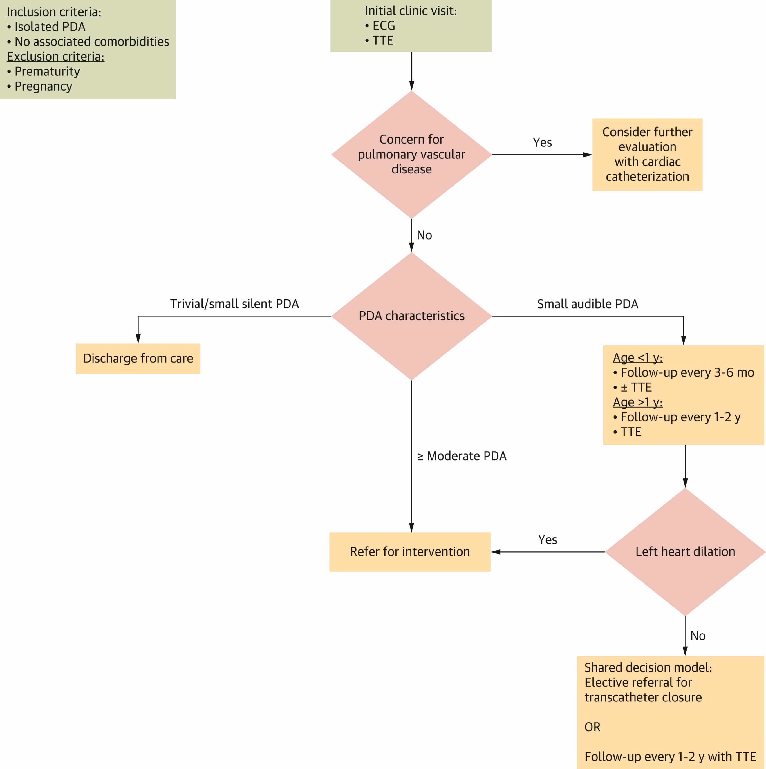
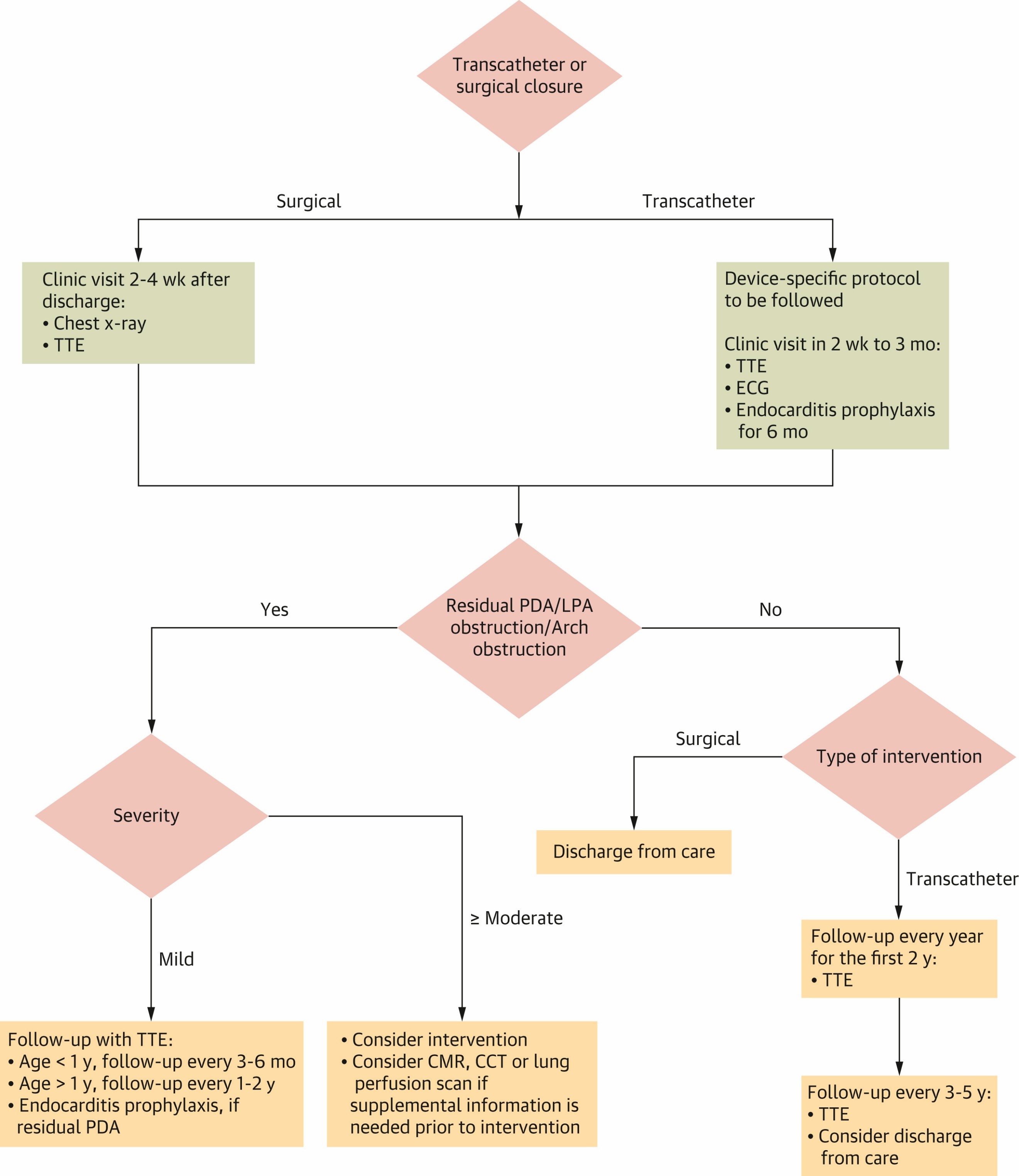
The goal of the PDA clinical practice algorithm is to provide cardiologists and other health care providers with a decision-support tool for clinical evaluation and follow-up testing of PDAs (Figures 1a, b). This guidance is not meant to replace health care providers' clinical judgment but rather to improve the value of care by reducing practice variation and optimizing resource utilization.
Figure 1a: Outpatient Management of Isolated PDA
Reprinted with permission from Writing Committee, Sachdeva R, Parthiban A, et al. Outpatient management of isolated left-to-right shunt lesions in pediatric patients: 2026 ACC concise clinical guidance: a report of the American College of Cardiology Solution Set Oversight Committee. J Am Coll Cardiol. Published online March 26, 2026. doi:10.1016/j.jacc.2025.11.020
ECG = electrocardiogram; PDA = patent ductus arteriosus; TTE = transthoracic echocardiography.
Figure 1b: Outpatient Management of PDA Post Intervention
Reprinted with permission from Writing Committee, Sachdeva R, Parthiban A, et al. Outpatient management of isolated left-to-right shunt lesions in pediatric patients: 2026 ACC concise clinical guidance: a report of the American College of Cardiology Solution Set Oversight Committee. J Am Coll Cardiol. Published online March 26, 2026. doi:10.1016/j.jacc.2025.11.020
CCT = cardiovascular computed tomography; CMR = cardiovascular magnetic resonance; LPA = left pulmonary artery; PDA = patent ductus arteriosus; TTE = transthoracic echocardiography.
The algorithm provides guidance for outpatient management before and after intervention. Each part starts with the initial standard workup and progresses to follow-up frequency and testing on the basis of PDA size and hemodynamic significance. In the case of the trivial, silent PDA, the algorithm recommends discharge from care.3,4 Frequency of follow-up and testing with echocardiography in various clinical scenarios are guided by the 2020 ACC/AHA/Multisociety Appropriate Use Criteria for Multimodality Imaging During the Follow-Up Care of Patients With Congenital Heart Disease and the 2026 ACC concise clinical guidance on Outpatient Management of Isolated Left-To-Right Shunt Lesions in Pediatric Patients.4,5 Following a small PDA infrequently rather than discharging from care is considered reasonable, given in some cases that it may result in left heart dilation over time, which would be an indication for intervention. The postintervention algorithm addresses follow-up on the basis of residual PDA, left pulmonary artery obstruction, and arch obstruction.3,6 In the case of surgical PDA closure, the patient may be discharged from cardiology care if there are no concerns at the postoperative clinic visit. Following transcatheter PDA device closure, annual follow-up for the first 2 years with echocardiogram is recommended. If there are no concerns, the clinician may consider discharge or follow-up every 3-5 years on the basis of institutional or device protocol.4,7
(For management of unrepaired atrial septal defects [ASDs] in patients >18 years of age, refer to the 2025 ACC/AHA/Multisociety Guideline for the Management of Adults With CHD.8)
Methods: Algorithm Development
The PDA clinical practice algorithm was developed by the Quality Working Group of the American College of Cardiology (ACC) Adult Congenital and Pediatric Cardiology (ACPC) member section. This algorithm was developed in a manner similar to the secundum ASD algorithm using the existing guidelines and the 2026 ACC concise clinical guidance on left-to-right shunt lesions.4,5,9
Future Directions
The PDA clinical practice algorithm can be integrated by clinicians as a decision-support tool in their practices. Furthermore, centers can use them for quality-improvement initiatives. The widespread use and implementation of the PDA algorithm could improve resource utilization and standardize the care of patients with PDAs.
References
- Romans RA, Rockefeller TA, Hancock HS. The physiologic implications of congenital heart defects. Semin Pediatr Surg. 2021;30(2):151042. doi:10.1016/j.sempedsurg.2021.151042
- Hokanson JS, Ring K, Zhang X. A survey of pediatric cardiologists regarding non-emergent echocardiographic findings in asymptomatic newborns. Pediatr Cardiol. 2022;43(4):837-843. doi:10.1007/s00246-021-02795-8
- Feltes TF, Bacha E, Beekman RH 3rd, et al. Indications for cardiac catheterization and intervention in pediatric cardiac disease: a scientific statement from the American Heart Association. Circulation. 2011;123(22):2607-2652. doi:10.1161/CIR.0b013e31821b1f10
- Sachdeva R, Valente AM, Armstrong AK, et al. ACC/AHA/ASE/HRS/ISACHD/SCAI/SCCT/SCMR/SOPE 2020 appropriate use criteria for multimodality imaging during the follow-up care of patients with congenital heart disease: a report of the American College of Cardiology Solution Set Oversight Committee and Appropriate Use Criteria Task Force, American Heart Association, American Society of Echocardiography, Heart Rhythm Society, International Society for Adult Congenital Heart Disease, Society for Cardiovascular Angiography and Interventions, Society of Cardiovascular Computed Tomography, Society for Cardiovascular Magnetic Resonance, and Society of Pediatric Echocardiography. J Am Coll Cardiol. 2020;75(6):657-703. doi:10.1016/j.jacc.2019.10.002
- Writing Committee, Sachdeva R, Parthiban A, et al. Outpatient management of isolated left-to-right shunt lesions in pediatric patients: 2026 ACC concise clinical guidance: a report of the American College of Cardiology Solution Set Oversight Committee. J Am Coll Cardiol. Published online March 26, 2026. doi:10.1016/j.jacc.2025.11.020
- Markush D, Tsing JC, Gupta S, et al. Fate of the left pulmonary artery and thoracic aorta after transcatheter patent ductus arteriosus closure in low birth weight premature infants. Pediatr Cardiol. 2021;42(3):628-636. doi:10.1007/s00246-020-02523-8
- Reo R, Van Pelt E, Lovelace C, et al. Utility of follow-up echocardiograms in uncomplicated PDA device closures performed after infancy. Cardiol Ther. 2022;11(3):445-452. doi:10.1007/s40119-022-00272-8
- Gurvitz M, Krieger EV, Fuller S, et al. 2025 ACC/AHA/HRS/ISACHD/SCAI guideline for the management of adults with congenital heart disease: a report of the American College of Cardiology/American Heart Association Joint Committee on Clinical Practice Guidelines. J Am Coll Cardiol. 2026;87(7):822-976. doi:10.1016/j.jacc.2025.09.006
- Sachdeva R, Parthiban A, Birnbaum B, et al. Clinical Practice Algorithm For the Follow-up of Unrepaired and Repaired Secundum Atrial Septal Defects. 2026. Available at: https://www.acc.org/Latest-in-Cardiology/Articles/2022/03/08/19/34/ Clinical-Practice-Algorithm-For-the-Follow-up-of-Unrepaired-and-Repaired-SASD. Accessed 08/2026.
Clinical Topics: Arrhythmias and Clinical EP, Cardiovascular Care Team, Congenital Heart Disease and Pediatric Cardiology, Diabetes and Cardiometabolic Disease, Heart Failure and Cardiomyopathies, Noninvasive Imaging, Prevention, Pulmonary Hypertension and Venous Thromboembolism, SCD/Ventricular Arrhythmias, Atrial Fibrillation/Supraventricular Arrhythmias, Congenital Heart Disease, CHD and Pediatrics and Arrhythmias, CHD and Pediatrics and Imaging, CHD and Pediatrics and Prevention, CHD and Pediatrics and Quality Improvement, Pulmonary Hypertension, Echocardiography/Ultrasound, Hypertension
Keywords: Ductus Arteriosus, Patent, Aftercare, Follow-Up Studies, Goals, Hypertension, Pulmonary, Consensus, Dilatation, Outpatients, Patient Discharge, Pulmonary Arterial Hypertension, Pulmonary Artery, Quality Improvement, Heart Septal Defects, Atrial, Hemodynamics, Echocardiography, Vascular Resistance, Electrocardiography, Ambulatory Care, Tachycardia, Morbidity, Algorithms, Tachypnea
< Back to Listings