18F-FAPI-42 PET Detects Fibroblast Activation in Myocardium in Patients With DCM
18F-labeled fibroblast activation protein inhibitor tracer (18F-FAPI-42) PET imaging can detect fibroblast activation in the myocardium among patients with dilated cardiomyopathy (DCM), according to a study published July 30 in JACC: Cardiovascular Imaging.
Sihao Liang, MM, et al., report on 19 patients with DCM who underwent 18F-FAPI PET/CT imaging, 14 of whom subsequently underwent cardiac PET/cardiac magnetic resonance (CMR). Control groups were used to identify a normal range for both imaging modalities.
In patients with DCM, the authors found that FAPI uptake in the myocardium was “intense and diffuse,” with varying degree of uptake across different regions. The control group did not exhibit radio-tracer uptake. Of note, more abnormal segments were identified with 18F-FAPI PET (n=168) than with CMR imaging (n=95). Plus, elevated T1-postcontrast values, greater extracellular volume %, and more severely impaired myocardial long-axis peak strain % capacity was present in FAPI-positive segments vs. FAPI-negative ones.
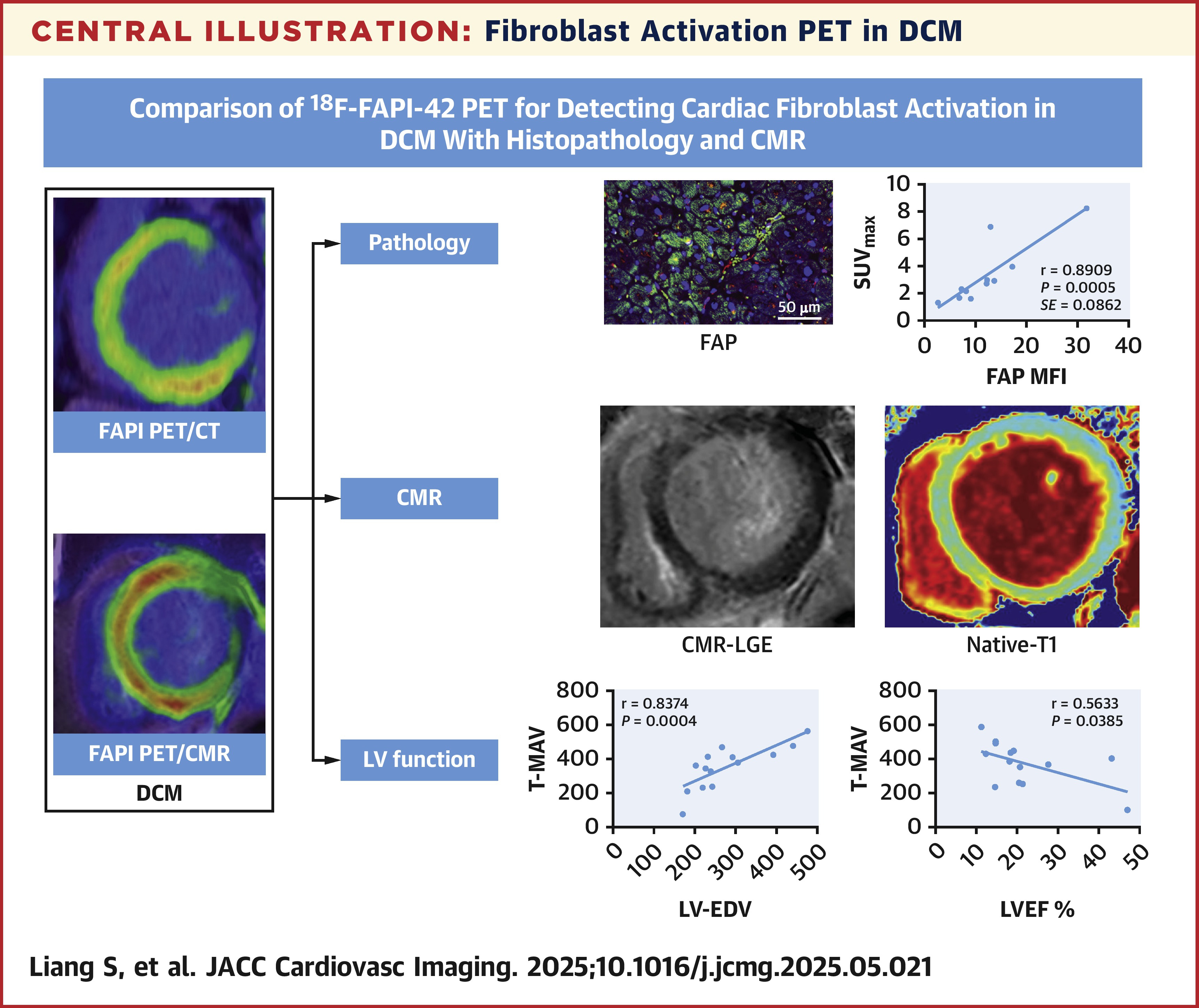
Within nine to 124 days after imaging, four study participants underwent cardiac transplantation. Among these patients, there was a strong correlation with FAPI uptake and FAP mean fluorescence intensity (p<0.001) and collagen fiber deposition (p<0.05). Associations were also noted between FAPI uptake and several parameters of cardiac function measured by CMR, including end-systolic volume, end-diastolic volume, LVEF %, and extracellular volume %.
The authors uncovered a consistent trend where metabolically active volume increases with progressively worsening NYHA functional class; however, myocardial FAPI uptake initially increased but then proceeded to decline.
“The correlation between [metabolically active volume] and CMR-derived cardiac function indicators and echocardiographic parameters was stronger than that of other semiquantitative parameters,” write the authors. “These findings suggest that [metabolically active volume] is an effective indicator for monitoring disease progression in DCM patients.”
Clinical Topics: Heart Failure and Cardiomyopathies, Invasive Cardiovascular Angiography and Intervention, Noninvasive Imaging, Interventions and Imaging, Computed Tomography, Nuclear Imaging
Keywords: Positron Emission Tomography Computed Tomography, Cardiomyopathy, Dilated, Positron-Emission Tomography, Magnetic Resonance Spectroscopy, Myocardium, Fibroblasts
< Back to Listings