Cutting-Edge Structural Interventions | Innovations to Reduce Radiation Exposure For Interventionalists
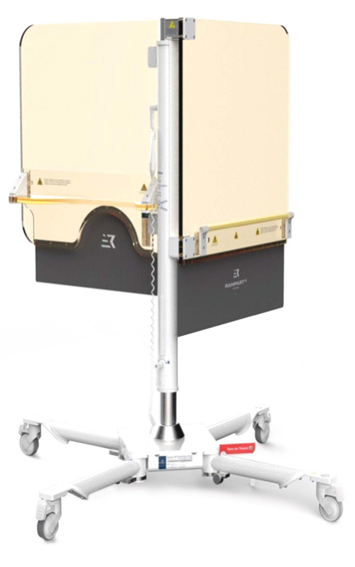
Figure 1: The Rampart M1128 Radiation Protection System
Most diagnostic and therapeutic cardiovascular procedures utilize ionizing radiation to image the heart. Despite the enormous benefit of these techniques on cardiovascular outcomes, ionizing radiation can be harmful to both patients and medical professionals. While the risk to patients is low (given relatively infrequent lifetime exposure), medical personnel are at a higher risk given their prolonged and frequent exposure to radiation.
Radiation exposure during invasive cardiovascular procedures remains at the upper range for diagnostic studies. It is even higher in complex electrophysiologic, coronary and structural heart interventions. The potential consequences of this exposure have been categorized for individuals, populations and occupationally exposed health care workers.
Lifetime doses of radiation more than 100 mSv are rare for patients but associated with an increased cancer risk. For comparison, the background radiation exposure for people living in the U.S. is about 3 mSv/y. When multiple members of a population are exposed, there is a potentially higher incidence of cancer and sequelae of radiation exposure.
Importantly for the cardiovascular community, physicians and health care staff may receive exposure of 10 mSv/year over a >30-year career. The implications of this level of radiation exposure have never been specifically studied, but multiple reports exist of radiation-associated eye lens damage increasing risk of cataracts, DNA damage, and increased rates of glioblastoma. Notably, the dose threshold to develop a malignancy is unknown as a single area of DNA damage can create an oncogene.
While radiation safety training is required at every institution, the concepts can be difficult to understand, the definitions of exposure can be confusing, and mitigation strategies lack significant innovation. Next time you walk out of a case, ask another team member how much radiation they were exposed to – chances are you'll get a blank stare. Let's start by simplifying!
The ACC has defined terminology surrounding radiation exposure for diagnostic and interventional procedures as follows:
- Exposure
- The amount of radiation that impinges on tissue
- Absorbed Dose
- The concentration of energy deposited by radiation in a specific tissue
- Measured in Gy (1 Gy = 1 Joule/kg of tissue)
- Equivalent Dose
- The absorbed dose adjusted by a weighting factor that accounts for the degree of biological damage caused by various types of radiation
- Measured in Sv
- Photons (x-rays and gamma rays) have a weighting factor of 1, therefore absorbed dose and equivalent dose are the same value
- Effective Dose
- The entire-body quantity used to compare effect from radiation on an average subject from a particular exposure
- Measured in mSv
- X-ray Fluoroscopic Air Kerma
- The level of radiation present at a specific location
- Measured in Gy
Radiation in cardiovascular imaging consists of photons and positrons. When these interact with body tissues, electrons are ejected from atoms forming ions and free radicals – this causes molecular damage. X-rays commonly used in fluoroscopy suites are transmitted by photons. Air kerma is the standard unit of measure for x-ray beam exposure.
Strategies to Mitigate Radiation Exposure
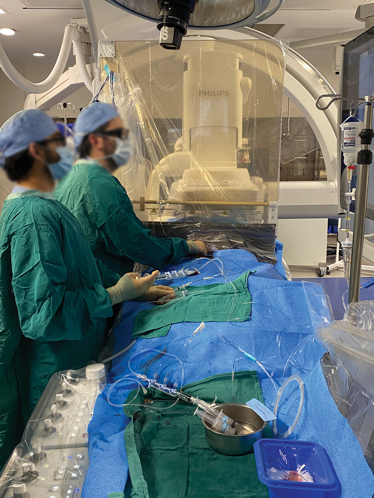
Figure 2: Performing a procedure while using the Rampart M1128 radiation protection system.
Traditionally, lead aprons, humeral shields, eye protection and blocking equipment have been used to mitigate the cardiovascular care team's radiation exposure. Standard lead aprons with a thyroid collar protect the axial skeleton (often leaving arms and hands vulnerable), cervical bone marrow and radiosensitive thyroid. Lead (0.5 mm) absorbs 95% of 70 kVp x-rays and 85% of 100kVP x-rays. Lead hats may provide an additional layer of protection that to date is largely theoretical.
Current apron and lead blocking equipment are designed to shield proceduralists and laboratory staff from radiation. However, previous studies have documented that orthopedic injuries and musculoskeletal pain are a likely result of wearing heavy leaded aprons. A large, multicenter study found that interventional laboratory employees reported a 67% increase in the prevalence of musculoskeletal pain.
Two Society of Cardiovascular Angiography and Intervention (SCAI) surveys were performed a decade apart in 2004 and 2014, both showing similar results for the orthopedic complications of the operators who wear leaded apron protection. The surveys revealed a 60% incidence of orthopedic issues in operators who have been working more than 20 years in the cath lab compared with a 2.3% incidence of chronic spine problems in the general population. Interestingly, of the operators who had been in practice less than five years, 26% already had orthopedic complaints.
The study also revealed that more than a third of those surveyed already had to take spine-related periods of absence from the cath lab. These injuries significantly impact staff satisfaction, patient throughput and lab productivity.
Innovation is necessary to overcome these limitations. The ideal shielding system is portable and adjustable.
The Zero-Gravity radiation protection system consists of a suspended 1.0 mm lead body shield and a 0.5 mm lead equivalent acrylic face shield to protect the head, eyes and neck of the operator. The system is designed to eliminate the weight burden of traditional heavy lead apparel for the operator with the freedom of movement.
In a study evaluating the efficacy of suspended stems, it was shown to provide superior radiation attenuation for operators compared with conventional lead apron shielding. A limitation of this suspended stem system is that the protection is only afforded to individuals operating the system – not the entire cath lab team.
Although focus on individual radiation attenuation technologies that support both attenuation and orthopedic safety have evolved, the emphasis has been on the individual operator. Until recently, a solution has not existed to address radiation attenuation and orthopedic safety for the entire scrubbed interventional team with one system.
To address the challenge of radiation protection in this environment without the burden of wearing a heavy lead apron, the Rampart, M1128 was developed. It is a fully adjustable and portable system that provides radiation protection for operators and their team without having to wear a lead apron.
Rampart M1128 is a configurable, floor-supported, portable shielding system with recommended under table lead (x 2) and one abdominal protector (Figure 1). This portable shielding system has floor casters to allow for easy movement and is placed on the right side of the procedure table. The system has two panels (each 1 mm thick Pb – twice the thickness of traditional wearable lead), a center mast and two Rampart Accessory Kits with soft shielding on each panel (0.5 mm thick Pb). A sterile plastic drape is used for each procedure to ensure sterility.
The design of the Rampart system allows the entire cardiovascular team on the right side of the patient to perform a case without wearing lead (Figure 2). The device is unique in that it allows for multiple vascular access points, protects not only the axial but also the appendicular skeleton, and adjusts for different table heights. Because the system does not isolate the primary operator, it allows for more traditional procedural ergonomics and mobility.
This system may represent the first step toward "lead free" invasive cardiovascular procedures and has been featured on live case transmissions during CTO Summit 2021 and CTO Live Aid.
Emory Cardiology has strongly supported staff radiation safety and well-being by acquiring multiple Rampart systems. After a one-week training, the faculty, fellows and staff develop proficiency in using the system. Added benefits for trainees are the ability to observe a procedure without "finding lead" nor do they need to struggle to find lead that fits appropriately.
To date, we have used the Rampart system during diagnostic coronary angiography, structural heart interventions (including LAMPOON and BASILICA), CTO PCI, and electrophysiologic procedures. We have a study underway to compare radiation exposure during elective cases that use the Rampart system.
Radiation safety remains an essential component of modern cardiology practice. New innovations have the potential to not only decrease the dangers of radiation exposure but also orthopedic injuries. Soon, the occupational hazards of procedural cardiology may be obsolete.
Cutting-Edge Structural Interventions is designed, in part, to highlight practice-changing science and technologies of interest to the interventional community. These pieces reflect the opinions of the author and are not endorsements of a product or service. Have a suggestion for a future topic or feature? Contact cardiologyeditor@acc.org.


This article was authored by John Lisko, MD, MPH; Nik Shekiladze, MD; and Pratik Sandesera, MD, interventional Fellows in Training at the Emory Structural Heart and Valve Center in Atlanta, GA.
Clinical Topics: Invasive Cardiovascular Angiography and Intervention, Noninvasive Imaging, Interventions and Imaging, Angiography, Nuclear Imaging
Keywords: ACC Publications, Cardiology Magazine, X-Rays, Electrons, Lead, Radiation Protection, Background Radiation, Incidence, Glioblastoma, Photons, Gamma Rays, Prevalence, Bone Marrow, Musculoskeletal Pain, Thyroid Gland, Plastics, Coronary Angiography, Percutaneous Coronary Intervention, Laboratories, Personal Satisfaction, Patient Satisfaction, Oncogenes, Fluoroscopy, Physicians, Health Personnel, Lens, Crystalline, Infertility, Cataract, Cardiology, Ions, Faculty, Free Radicals, DNA Damage, Reference Standards, Innovation
< Back to Listings