Cutting-Edge Structural Interventions | Building a Structural Program: Invest in Your Imagers

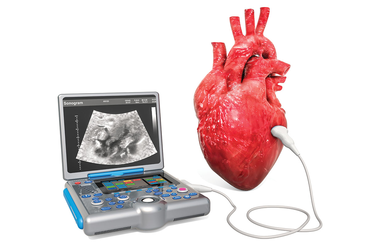
A key lesson I learned early when working to build a structural heart program is that we must be able to see severe valvular heart disease to diagnose it and ultimately treat it. Thus, the most valuable education you can provide is to your sonographers.
No matter how current referring physicians are with the latest guidelines and no matter how aggressive they may be with referring patients early in their disease progression, there won't be any referrals to your program without the ability to correctly identify valvular heart disease.
Perhaps the echocardiography laboratory in your institution is already doing well – in this case your job is a lot easier. But often there is room for improvement, and this may be more likely in a community hospital or a private practice setting. As you plan this education, don't go at it alone. Partner with your cardiac imaging specialist, whose assistance will be invaluable as you blaze a trail forward. Here I review key considerations in optimizing the imaging for aortic stenosis and mitral regurgitation to optimize imaging protocols and education for the team.
Imaging For Aortic Stenosis
Severe aortic stenosis (SAS) seems to be the most frequently diagnosed form of valvular heart disease. Unlike its counterpart, severe mitral regurgitation, SAS seems straightforward to diagnose. Patients who meet distinct criteria (usually a maximum velocity ≥4 meters/second or mean gradient ≥40 mm Hg across the aortic valve) are readily diagnosed with SAS, assuming the aortic valve looks diseased/restricted and there is no sub- or supravalvular obstruction.
Clearly, surgical or transcatheter aortic valve replacement (SAVR, TAVR) is lifesaving for patients diagnosed with SAS. Now, the group of patients who can benefit from TAVR has expanded in recent years to those who do not meet these classic diagnostic thresholds.1-4 In such patients, dobutamine stress echocardiography (DSE) has been heralded as the historical gold standard; yet challenges with this test limit its use in clinical practice.5
In my own practice, after experiencing firsthand the futility of resuscitating a coding patient with SAS in the echocardiography laboratory, I find it difficult to justify conducting a DSE on patients with probable SAS. There are scant data as to the safety of DSE for these patients and usually the risks do not outweigh the benefits, especially when we have viable alternatives. Also, DSE is a resource-intensive test, potentially requiring an hour or more to perform. Thus, DSE is challenging to schedule in a community hospital and usually requires a provider to be present the entire time. Furthermore, the utility of DSE in patients with paradoxical low-flow, low-gradient SAS with preserved ejection fraction is questionable and may lead to harm.
Therefore, DSE is usually underutilized; hence, aortic stenosis is often underdiagnosed and undertreated. Many patients who could potentially benefit from TAVR or SAVR are never identified. There are three alternatives to DSE: 1) 2D aortic valve planimetry by transesophageal echocardiography (TEE), 2) aortic valve calcium scoring by computed tomography, and 3) use of contrast-enhanced ultrasound agents (CEUS) during transthoracic echocardiography (TTE).
Aortic Valve Calcium Scoring
Aortic valve calcification as measured by computed tomography is an accurate, reproducible and well-validated marker of stenosis severity and disease progression, as well as a powerful predictor of adverse events. Although Doppler echocardiography is used for first-line evaluation of the severity of AS, resting echocardiographic assessment is discordant in up to 40% of patients, leading to clinical uncertainty.6
Interest has grown, therefore, in aortic valve calcium scoring using multidetector computed tomography (CT-AVC) as an alternative assessment of AS severity. Measurement of CT-AVC requires a gated, noncontrast CT scan. Sex-specific thresholds of 1,200-1,300 Agatston units (AU) for women and 2,000 AU for men should be applied to identify SAS.7
At my institution, our use of CT-AVC as a diagnostic tool has been limited by the challenge of obtaining insurance approval. Although we offer testing for coronary artery calcium scores, there's a poor track record of obtaining reimbursement from many of the major insurers, which consider this testing to be experimental. Instead, as a workaround, if a patient is to undergo cardiac CT angiography (CTA) with contrast, we add a gated study without contrast to the test protocol, which allows the clinician to evaluate the patient for possible AS. This requires close collaboration with the department responsible for cardiac CTA (radiology or cardiac imaging).
Another option is to include a noncontrast-gated cardiac CTA as part of the TAVR CTA acquisition protocol. Again, a discussion with your radiologist or imaging cardiologist should make this part of your protocol.
Contrast-Enhanced US Agents
Unlike red blood cells, which are poor scatterers of US, the microbubbles in CEUS agents are compressible and they have a different density than blood cells. This unique physical characteristic of microbubbles is important in order to understand the behavior microbubbles exhibit when exposed to ultrasound energy.8
The utility of CEUS agents to evaluate the severity of AS dates back to publications in the early 1990s.9
In a study in 2002, the role of CEUS Doppler echocardiography was compared with catheterization measurements as the gold standard in the assessment of AS severity. Contrast Doppler yielded higher peak gradients than conventional Doppler (85.6 +/- 30.2 vs. 72.6 +/- 26.1 mm Hg; p<0.001), as well as higher mean gradients (51.4 +/- 19.0 vs. 44.2 +/- 15.9 mm Hg; p<0.001). There was no difference between mean contrast Doppler gradients and mean catheterization gradients, which showed a high correlation (r=0.89; p<0.001).10
The current clinical recommendations from the European Association of Cardiovascular Imaging and the American Society of Echocardiography (ASE) state that in the case of "poor acoustic quality, the use of echo contrast media has been suggested but it is not used in many echocardiography laboratories. In case of its use, proper machine settings are crucial to avoid artifacts..."11
This document is 100% right. CEUS agents typically are not used in many echocardiography laboratories to assess the severity of AS. Thus, I would argue that patients with severe AS are often misdiagnosed with moderate AS.
I've had many patients in the past year who had classic, progressive symptoms of AS, including NYHA class 3 symptoms and multiple admissions for heart failure. These patients, despite having a preserved ejection fraction, still had a mean aortic valve gradient in the 25-30 mm Hg range and maximum velocity across the aortic valve of approximately 3.4 meters/second. However, on repeat echocardiography with CEUS agents, the mean gradient was >40 mm Hg and the patient then qualified for AVR.
After a discussion about this with our sonographers, many asked whether the CEUS agent was spuriously increasing the aortic gradients. Together we worked to answer this question. In patients with heavily diseased aortic valves and those with mildly sclerotic aortic valve disease who were being given a CEUS agent for another reason, the sonographers measured the aortic valve gradient before and after the CEUS injection. Their findings were striking.
In patients with mildly sclerotic aortic valves, CEUS agents across the board increased the measured gradients slightly, but never to a pathological degree. The sonographers could not find a single example of a patient in whom CEUS agents "gave" the patient a false diagnosis of SAS. Of course, this was hardly a scientific study. Yet, this finding convinced our sonographers of the importance of paying close attention to the aortic valve and lining up the Doppler cursor when investigating patients for SAS, as well as making use of CEUS agents in assessing patients with AS.
This small modification, over the past year, has allowed our sonographers to identify more patients with severe symptoms who meet the guideline criteria for SAS, who previously had been diagnosed with either moderate or moderate-to-severe AS. Moreover, this has reduced our utilization of other tests with much higher risk profiles, including DSE. Importantly, this has given more patients who would benefit the opportunity to be treated with an AVR.
It must be noted, however, that the use of CEUS in this setting requires study regarding its efficacy for clinical decision-making.
Imaging For Mitral Regurgitation
Unlike its next-door neighbor within the heart, the mitral valve can prove infinitely trickier to assess. Even those with mastery in cardiac sonography can be fooled at least once in their careers. Although I am board certified in adult echocardiography and feel very comfortable with assessing valvular regurgitation and stenosis on both TTE and TEE, I find myself soliciting second opinions from colleagues on studies with incongruent findings.
The main contributor to this difficult interpretation is that we're limited to only the data from the echo images obtained by the sonographer. Unlike with CT, where the raw data are available for review if needed by the interpreting physician, with sonography there's no additional data if, for example, a color or continuous wave Doppler was not performed with good technique. The only option is a repeat test, which is inconvenient for the patient and adds to the overall cost of care.
And adding to the complication of interpreting the echo images are the numerous quantitative and qualitative measures that can be obtained but that don't always agree with one another. A full review of the mitral valve by echocardiography is certainly beyond the scope of this article (and my word limit!) but interested readers should consult the latest ASE guidelines.12 However, I must emphasize the need for ongoing support and education for sonographers.
Prioritizing Education For Sonographers
In busy clinical practices, both at community and academic practices, sonographers and the entire CV team may not receive structured continuing education at their workplace. Our director of noninvasive and advanced cardiovascular imaging, Arash Seratnahaei, MD, FACC, established weekly and monthly education sessions for our sonographers. He created a formal curriculum for the sonographers, who earn continuing education credits by attending. The learning sessions are an open forum for sonographers to discuss interesting cases and ask questions along with staying up to date with lectures from our most experienced echocardiographers.
My cardiac anesthesiology partner, Oscar Penate, MD, and I gave a few lectures on mitral valve imaging, as well as reviewed the transcatheter edge-to-edge repair (TEER) procedure and things to look for on pre- and postoperative exams. This was immensely helpful for the sonographers, most of whom had never seen or scanned a patient who had undergone TEER, because the technology was new to our community.
Most importantly, we try to keep the sonographers current through constant feedback on the quality of studies, etc. I make it a point to congratulate my sonographers on studies that are done well and provide constructive feedback on studies that may have missed the mark. In this way, our sonographers are able to improve their own skill as well as the overall benchmark standard of the entire echocardiography laboratory.
Setting Priorities
When starting or growing a structural heart program, there are tremendous time demands for you and your team. Everyone will want to provide suggestions and a helping hand, but there are only so many hours in the day. Priorities must be made.
Make your sonographers a priority. As soon as I started at my institution, I made it a point to get to know my sonographers, including those who work at remote sites one or two hours away. All are invited to education and team-building sessions. Early on (and pre-COVID of course), I organized dinner events where they could learn about the new procedures we would be providing, including TEER and left atrial appendage occlusion. And we provided them training in performing intraprocedural TAVR imaging, something new to them. Most importantly I made sure I was easily accessible to the sonographers; I gave them my cell phone number and encouraged them to call or text anytime for any questions on studies.
Building and supporting the team – and being aware of the education needed and the partnership required to provide the best care for patients – will continue to pave the road to success on the quest to build and grow your structural heart program.

This article was authored by Sandeep Krishnan, MD, RPVI, FACC, an interventional and structural cardiologist and director of Structural Heart Disease at the King's Daughters Medical Center in Ashland, KY.
Do you have a topic or question to be addressed in this series? Tweet the author @SKrishnanMD or email him at xsandeepx@gmail.com. Thank you.
Clinical Topics: Cardiac Surgery, Cardiovascular Care Team, Heart Failure and Cardiomyopathies, Invasive Cardiovascular Angiography and Intervention, Noninvasive Imaging, Valvular Heart Disease, Aortic Surgery, Cardiac Surgery and Heart Failure, Cardiac Surgery and VHD, Acute Heart Failure, Interventions and Imaging, Interventions and Structural Heart Disease, Computed Tomography, Echocardiography/Ultrasound, Nuclear Imaging, Mitral Regurgitation
Keywords: ACC Publications, Cardiology Magazine, Anesthesiology, Aortic Valve, Aortic Valve Stenosis, Artifacts, Atrial Appendage, Benchmarking, Blood Cells, Calcium, Cardiologists, Catheterization, Cell Phone, Clinical Decision-Making, Constriction, Pathologic, Computed Tomography Angiography, Contrast Media, Coronary Vessels, Curriculum, Diagnostic Errors, Disease Progression, Echocardiography, Echocardiography, Doppler, Echocardiography, Stress, Echocardiography, Transesophageal, Education, Continuing, Erythrocytes, Feedback, Heart Failure, Hospitals, Community, Insurance Carriers, Laboratories, Meals, Medical Futility, Microbubbles, Mitral Valve, Mitral Valve Insufficiency, Physicians, Radiologists, Radiology, Referral and Consultation, Stroke Volume, Technology, Tomography, Tomography, X-Ray Computed, Transcatheter Aortic Valve Replacement, Uncertainty, Workplace
< Back to Listings

